Peptide Sequence to Structure: A Step-by-Step Workflow
Learn how to move from a peptide sequence to structure with prediction, confidence ranking and bench validation for laboratory research.
Pasting a peptide sequence into a structure predictor and accepting its highest-scoring model is a tempting shortcut. It's also an unreliable way to move from peptide sequence to structure, especially when the molecule is short, chemically modified, cyclic, or studied with a binding partner. The useful output isn't a definitive shape. It's a set of testable structural hypotheses, ranked by their chemistry, reproducibility, and agreement with measurements.
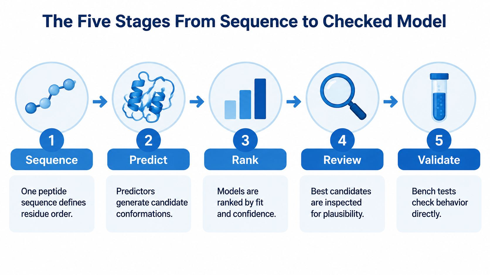
The Five Stages From Sequence to Checked Model
A peptide sequence defines residue order, but it doesn't necessarily define a single persistent shape. Solvent, temperature, concentration, chemical state, and binding partners can change which conformations are populated. A predictor may return a tidy structure even when the experimental sample contains an ensemble.
The distinction matters because protein-folding performance sets misleading expectations. AlphaFold2 achieved a median GDT_TS of 92.4 in CASP14, but that protein benchmark doesn't establish equivalent accuracy for short, flexible peptides, as the CASP14 assessment makes clear through its protein-focused results.
Workflow stages, outputs and pass checks
| Stage | What You Do | Output | Pass Check |
|---|---|---|---|
| Sequence preparation | Annotate termini, modifications, cyclization and binding partner | Annotated sequence record | Every residue and bond type is recorded |
| Prediction | Run a peptide-capable predictor with several seeds | Set of candidate models | Multiple seeds produce output |
| Ranking | Cluster poses and score them with confidence metrics | Shortlist of three to five poses | Top poses agree across seeds |
| Computational validation | Compare two predictors and check interface contacts | Confidence-ranked hypothesis | Agreement across independent methods |
| Experimental check | Measure the peptide with CD, NMR, HDX or a mutational assay | Measured data on the same peptide | Measurements match the predicted feature |
The stages separate different ways a model can fail. A structure can contain incorrect chemistry, propose the wrong receptor contacts, or represent a conformation that's scarcely populated under assay conditions.
More structural detail doesn't automatically mean more biological insight. The comparative analysis of peptide representations describes the difficulty of representing flexible peptides and notes that sequence-only baselines outperformed ensemble-aware representations on half of six downstream tasks. Ensembles can expose alternative conformations, while spectroscopy can test whether a proposed structural feature exists in solution.
Planning rule: Before prediction, the laboratory should record the peptide case, starting tool, and first check in a single line.
For example: a linear receptor-binding peptide, AlphaFold 3 complex prediction, then inspection of peptide–receptor contacts. That short plan keeps model generation connected to the scientific question rather than to the appearance of the output.
Prepare the Sequence and Match It to a Tool
The sequence record is the first experimental control in a computational workflow. A predictor can only model the molecular identity that its input describes.
- Confirm residue order and length. Check the sequence against synthesis documentation, not a shortened product name or an old spreadsheet.
- Mark noncanonical residues and D-amino acids. A standard amino-acid letter may not specify the intended stereochemistry or side-chain chemistry.
- Record both terminal states. Free, acetylated, amidated, and other capped termini aren't interchangeable.
- Specify every cyclization bond. Distinguish head-to-tail closure from side-chain or backbone-to-side-chain connections.
- List disulfides and modifications. Include connectivity, lipidation, and post-translational modifications rather than noting only that the peptide is modified.
- Name the binding partner. Record the intended receptor construct, chain boundaries, and relevant complex components.
A sequence notation isn't a complete molecular identity unless these details are captured. Residue polarity also informs inspection of solvent-facing and buried groups; the explanation of polar amino acids provides useful background without replacing explicit chemical annotation.
Match the chemistry before comparing tools
The review of cyclic-peptide prediction workflows describes improved prediction when cyclic positional constraints were introduced into AlphaFold2, with reduced accuracy when those constraints were removed. A terminal bond changes the modeling problem, not merely the final drawing.
Peptide case, starting tool and main caveat
| Peptide Case | Starting Tool | Main Caveat |
|---|---|---|
| Short linear peptide, no modifications | PEP-FOLD or ESMFold for a quick first pass | Flexible peptides can show several conformations |
| Cyclic peptide | AlphaFold 3 with the cyclic bond encoded, if supported | Cyclic constraints change accuracy substantially |
| Peptide with noncanonical residues | Rosetta with residue parameters for the modification | Chemistry must be encoded or results are unreliable |
| Peptide bound to a receptor | Rosetta FlexPepDock or AlphaFold 3 in complex mode | Interface geometry is less certain than the global fold |
| Lipidated or heavily modified peptide | Prediction as a hypothesis only, with experimental data first | No general benchmark validates these chemistries |
ESMFold can provide a provisional starting geometry for an ordinary linear sequence. It shouldn't be treated as confirmation of a solution structure or binding pose.
For unusual chemistry, the model and any subsequent force field must support the intended residues and bonds. If any annotation remains unresolved, the sequence isn't ready for prediction.
Read Confidence Scores Without Overtrusting Them
Confidence scores answer different questions. pLDDT describes local confidence around individual residues, whereas predicted aligned error describes uncertainty in the relative placement of residue pairs. Neither alone establishes that a peptide occupies the correct receptor-binding site.
Fnat measures the fraction of native-like interface contacts recovered against a reference. DockQ summarizes interface quality, while cluster support records whether repeated runs converge on a similar pose. Peptide backbone RMSD measures geometric deviation from a chosen reference after a clearly specified alignment.
Confidence metrics and how to read them
| Metric | What It Measures | How to Read It |
|---|---|---|
| pLDDT | Per-residue local confidence from 0 to 100 | Above 90 very high, 70 to 90 generally confident, 50 to 70 low, below 50 often disordered |
| Predicted aligned error | Confidence in the relative position of residue pairs | Low error between peptide and receptor residues supports the interface |
| Fnat | Fraction of native-like interface contacts | Higher values support the contact pattern; read alongside DockQ |
| DockQ | Overall interface quality from 0 to 1 | Below 0.23 incorrect; 0.23 to below 0.49 acceptable; 0.49 to 0.80 medium; above 0.80 high |
| Cluster support | Number of seeds converging on one pose | A majority of seeds in one cluster is stronger than a single top score |
| Peptide backbone RMSD | Deviation of the peptide backbone from a reference | Report separately from receptor RMSD |
Separate confidence from measured accuracy
A confident receptor fold can dominate the visual impression of a complex while the peptide is misplaced. Induced fit and context-dependent binding make that distinction particularly important.
Low pLDDT can be consistent with disorder, but it isn't an experimental disorder measurement. Likewise, low predicted aligned error supports a proposed relationship between chains without proving that the interaction occurs.
A reference-based score requires a reference. Without an experimental complex, true DockQ and Fnat aren't available merely because a prediction contains an interface.
Predicted interface scores must therefore be labeled as predictions rather than substituted for measured contact recovery. RMSD also needs an alignment definition: fitting the receptor before measuring peptide displacement answers a different question from fitting the peptide itself.
A pose should advance only when interface-focused assessments agree and its cluster recurs across seeds. A high local-confidence score can support that decision, but it shouldn't make the decision alone.
Run Predictions and Rank the Candidate Models
Prediction should produce a comparison set, not a single image selected because it looks plausible. Each run needs enough recorded context to explain why one candidate outranks another.
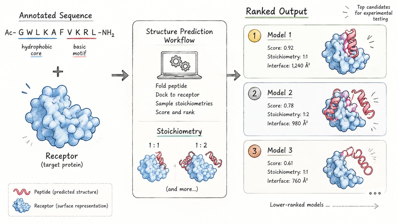
- Define the complex composition. Test biologically motivated peptide-to-receptor stoichiometries, such as 1:1 and 1:2, rather than assuming a chain count. Extra or missing chains can alter accessible surfaces and proposed contacts.
- Generate repeated predictions. A practical starting design is at least five random seeds per setting. This is a workflow choice, not a guarantee of adequate sampling; unresolved variability calls for broader exploration.
- Cluster by peptide backbone geometry. Use a consistent alignment and backbone RMSD definition. The aim is to distinguish recurring binding modes from small variations within the same mode.
- Rank clusters using complementary evidence. Combine interface contacts, peptide–receptor predicted aligned error, and local confidence. Where a reference exists, apply Fnat and the DockQ quality bands rather than treating a global fold score as an interface score.
- Inspect the leading poses in PyMOL or ChimeraX. Review the top three to five candidates for clashes, broken chemistry, implausible chain crossings, and sensible interactions. Hydrophobic side chains facing a compatible hydrophobic patch are more plausible than equivalent groups buried against unsupported polar contacts.
- Save a reproducible ranking record. Include tool version, inputs, seeds, stoichiometry, cluster membership, and rejection reasons. A rejected pose can be informative if a later experiment contradicts the preferred cluster.
The prediction and docking workflow should preserve alternative poses until evidence distinguishes them.
Interpret the shortlist at the interface
In a benchmark of 80 peptide–protein complexes, average TM-scores of approximately 0.91–0.92 accompanied average DockQ scores around 0.63, according to the peptide-complex prediction analysis. Strong overall geometry can therefore coexist with less certain interface placement.
A usable computational result has a dominant cluster that recurs across seeds and proposes chemically coherent contacts. Where binding residues are already known, agreement with those residues adds a separate constraint.
If competing clusters remain similarly supported, the ranking should preserve that ambiguity. Selecting a visually attractive pose doesn't resolve it; an experiment designed to distinguish the competing contacts might.
Catch Overfitting and Benchmark Pitfalls
A model can appear accurate because a test structure, or a close relative, was already represented during training. Short peptide benchmarks are particularly vulnerable when closely related sequences or complexes are counted as independent tests.
The bioRxiv benchmark analysis, a preprint, reports that removing overlapping cases reduced success rates to approximately 40–65% under a stringent contact-accuracy criterion. It also reports sub-1-Å RMSD for AlphaFold 3 on 90 of 394 targets, compared with 34 for AlphaFold 2, and residue-level RMSDs reaching 6.670 Å for noncanonical cyclic peptides, without a significant linear relationship between confidence and structural deviation.
Those findings describe different tests, not a universal success rate. Better performance on a broad peptide benchmark doesn't establish accuracy for every modified, cyclic, stapled, or disordered molecule.
Audit the comparison before accepting the gain
- Check structural overlap. Identify duplicated complexes, closely related receptor constructs, and peptide variants that differ only superficially.
- Separate homologous families. Build a low-homology holdout set rather than relying only on random splitting.
- Check release timing. Establish whether the evaluated structures could have been available during model development.
- Match chemistry to the claim. A benchmark dominated by canonical linear peptides cannot validate noncanonical cyclic chemistry.
- Keep the evaluation rule fixed. Changing the success criterion between methods prevents a meaningful comparison.
The relevant test is whether an apparent gain survives removal of overlapping or closely related cases. A laboratory evaluating a method should retain those exclusions in its methods record, including the criteria used to identify related structures.
Benchmark question: Would this pose still rank first if the nearest training homologue were removed?
That question may not be answerable for a closed training set. When training provenance is incomplete, the report should state the limitation rather than convert an unknown into an assumption of independence.
For chemically unusual peptides, unsupported confidence should lead to a targeted validation plan. It shouldn't lead to stronger wording in the structural conclusion.
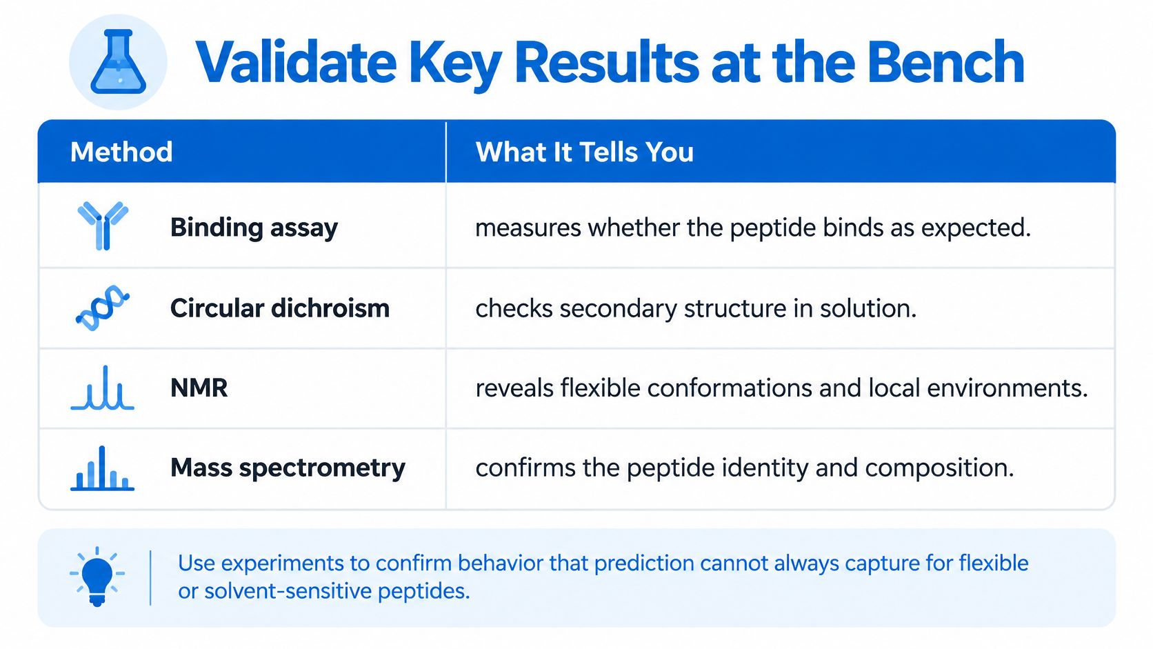
Validate Key Results at the Bench
Experiments measure the peptide's behavior under defined conditions. They can distinguish a persistent helix from a flexible ensemble, test whether a proposed interface matters, or reveal that a predicted feature disappears in the assay buffer.
The experimental method should match the structural claim. Careful assay development also separates a genuine structural effect from changes in solubility, aggregation, or readout performance.
Experimental methods and what each resolves
| Method | What It Resolves | Sample Requirement |
|---|---|---|
| Circular dichroism (CD) | Secondary structure content in solution | Dilute, buffer-compatible solution |
| NMR | Residue-level structure and dynamics | Soluble sample at suitable concentration |
| Hydrogen–deuterium exchange (HDX-MS) | Solvent exposure and binding-induced protection | Solution sample in an MS-compatible buffer |
| X-ray crystallography | Atomic structure of a crystallized complex | Crystallizable sample, often the hardest step |
| Cryo-EM | Near-atomic structure of larger complexes | Larger peptide–protein complexes, not small peptides alone |
| Mutational binding assay | Whether predicted interface residues are required | Mutant peptide variants and a binding readout |
For flexible peptides, molecular dynamics can explore alternative conformers before bench work. Its trajectories remain computational hypotheses, not measurements of conformer populations.
Preserve sample identity through preparation
The laboratory's validated protocol governs solvent selection and handling. The following routine concerns analytical or in-vitro sample preparation, not human administration.
- Equilibrate sealed vials before opening. The laboratory handling guide specifies approximately 20–30 minutes at room temperature to reduce condensation risk.
- Introduce compatible solvent slowly down the vial wall. Directing solvent onto the lyophilized cake can complicate controlled wetting and mixing.
- Mix gently. Gentle swirling is preferable to vigorous vortexing, foaming, or aggressive sonication, which can promote adsorption, aggregation, or structural disruption.
- Record lot-specific characterization. Identity by mass spectrometry, chromatographic purity, water content, and counterion information belong in the assay record.
ProTecPep does not provide dosing, administration, reconstitution, treatment, or medical-use guidance. Laboratory personnel remain responsible for their validated sample-preparation procedures.
Finally, compare the observation with the feature the experiment was designed to test: secondary structure, solvent protection, or loss of binding after mutation. After assay controls are checked, a mismatch requires revision of the model, not dismissal of the measurement.
Decide Which Model Is Ready to Report
A predicted peptide structure is a conformational hypothesis, never a measured structure. Support requires recurring clusters, interface agreement across independent predictors, and an experimental observation consistent with the specific proposed feature.
The reporting decision follows a defined sequence:
- Confirm recurrence. Establish that the preferred cluster appears across the seed set.
- Confirm interface agreement. Compare two independent predictors using appropriate interface assessments.
- Match an observation. Identify a bench result consistent with the proposed helix, contact, or binding loss.
- Assign the status. Label the hypothesis supported, partially supported, or unvalidated.
A cyclic, modified, or highly flexible peptide without an experimental check remains unvalidated, regardless of its scores. Partial agreement should be described at the feature level rather than extended to the whole structure.
Every report needs the tool and version, seed count, stoichiometry, scoring metrics, alignment definitions, and experimental methods. Physical samples also require identity and lot documentation; peptide purity testing supports that record but doesn't establish conformation.
ProTecPep does not warrant or guarantee any particular research outcome, experimental result, or scientific conclusion. Permitted work includes analytical testing, chemical characterization, method development, reference-standard comparison, stability testing, instrument calibration, in-vitro investigation, and other lawful non-clinical research.
A supported hypothesis describes agreement between a model and specified observations. It isn't blanket proof of activity, binding, or stability under untested conditions.
ProTecPep supplies research compounds intended exclusively for laboratory research, analytical testing, and scientific investigation. Researchers planning a peptide sequence-to-structure workflow can visit Pro Tec Pep to review research-compound information, then match the selected material's identity and documentation to the computational model and planned experimental check.