Peptide Sequence Determination: A Practical Lab Guide
Peptide sequence determination explained: MS/MS fragmentation, Edman degradation, sample prep and how to verify results in the lab.
The familiar advice to run a peptide through mass spectrometry and accept the best sequence match skips the most important work. Peptide sequence determination is an evidence-based assignment, not a direct readout. An analyst must connect measured masses to residue order, identify gaps in that evidence, and decide which additional experiment could resolve them.
A useful result therefore contains more than an amino-acid string. It explains what was measured, which residues are supported, and what remains uncertain. That distinction matters whether a laboratory is confirming a synthetic peptide or investigating an unexpected fragment.
Choosing a Method That Fits Your Peptide Sample
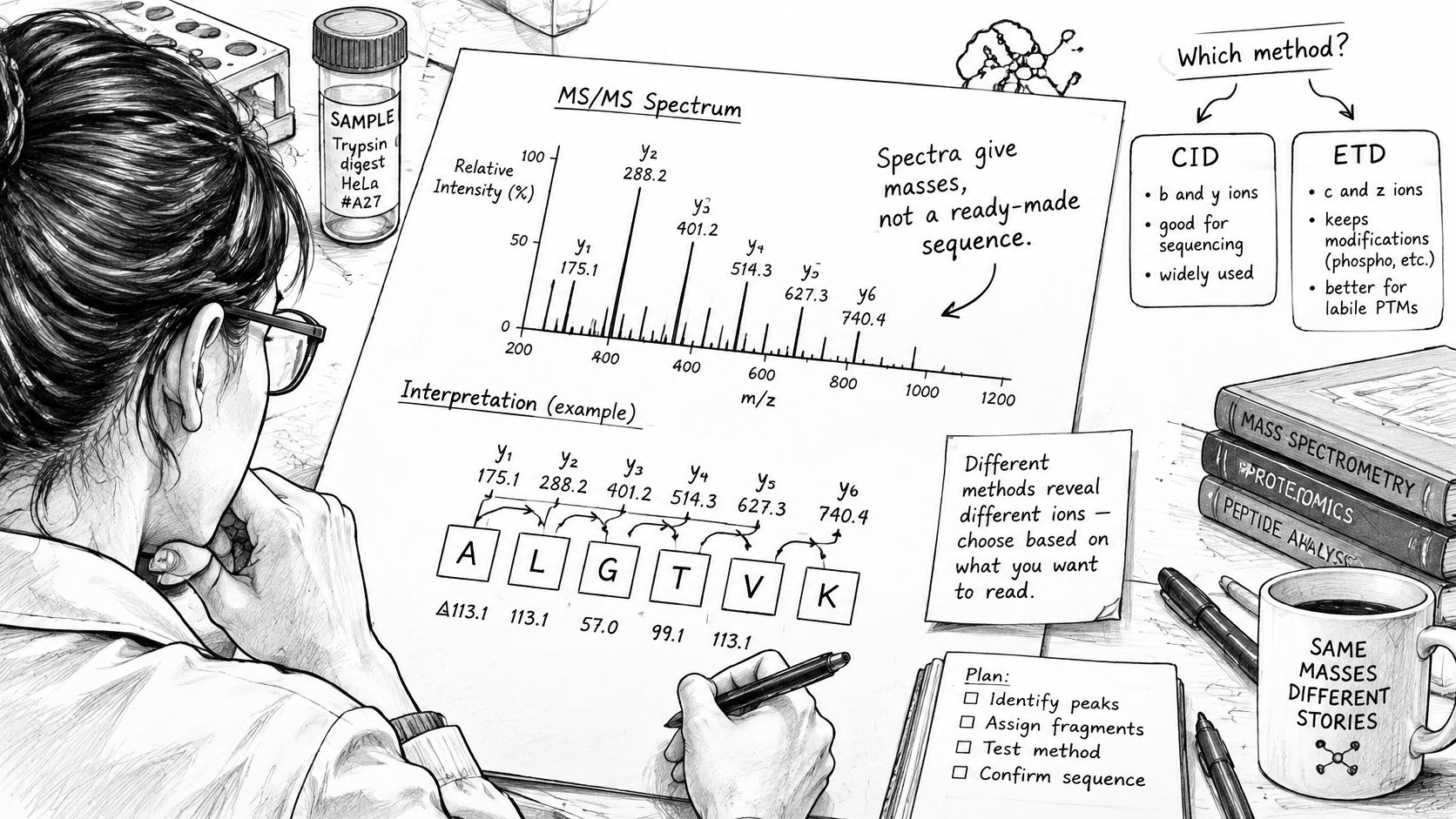
A mass spectrum provides masses that require interpretation, and the method chosen determines which parts of the sequence can be examined.
The starting point is the sample, not the available software. A purified short peptide, an intact protein, and a complex digest present different analytical problems. The comparison below separates the measurement each method provides from the conclusion an analyst might draw.
Sequencing methods by sample type and limit
| Method | Best for | Practical limit | Output |
|---|---|---|---|
| Edman degradation | Purified peptide with free N-terminus | About 30 residues routine readout | N-terminal order only |
| Database-search MS/MS | Peptide expected in a sequence database | Limited to sequences represented in the search space | Evidence supporting known peptide assignments |
| De novo MS/MS | Novel or unexpected peptide with no database match | Limited by fragment coverage | Order rebuilt from fragment mass gaps |
| Bottom-up LC-MS/MS | Complex mixtures after enzymatic digestion | Gaps where useful cleavage products are missing; may need multiple proteases | Sequence fragments for reconstruction |
| Top-down MS | Intact protein or large peptide | About 60% sequence coverage in current practice | Intact mass with partial sequence |
The top-down estimate and the need for complementary digestion strategies are discussed in the preprint on sequence-coverage limitations and complementary workflows. That coverage estimate describes current practice, not a guaranteed result for every sample.
A short decision path
- Is the peptide pure? A dominant purified species makes direct N-terminal analysis feasible. An unresolved mixture generally favors separation coupled to MS/MS.
- Is the N-terminus free? An accessible terminus supports Edman analysis. A blocked terminus directs attention toward mass-spectrometric methods.
- Is the peptide expected in a database? A represented sequence supports database searching. An unexpected sequence calls for de novo interpretation or an expanded search strategy.
- Is the sample a mixture? A protein mixture usually favors bottom-up LC-MS/MS. An intact-protein question may instead justify top-down analysis.
The analytical setting typically involves interpreting spectra alongside sample-preparation information.
Sequence determination also differs from determining molecular shape. The relationship between peptide sequence and structure helps define whether an experiment needs primary-sequence evidence, structural characterization, or both.
A laboratory worksheet should record the answers to these questions and the corresponding starting method. That choice remains provisional until the sample produces interpretable evidence.
Sample Preparation Choices That Change the Result
A preparation problem can resemble a sequencing problem. Salts, detergents, and buffer components can suppress ionization, introduce interfering signals, or obscure a peptide that is present in the vial. Increasing acquisition time won't necessarily correct unsuitable sample chemistry.
Cleanup should remove interference without selectively losing the analyte. Reversed-phase desalting can separate retained peptides from soluble salts, but a very hydrophilic peptide may not retain well. Detergent-containing samples may require a detergent-removal strategy rather than a desalting step alone.
Cleanup and digestion solve different problems
Cleanup improves compatibility with the instrument. Digestion changes the molecules being measured.
Trypsin cleaves after lysine and arginine under suitable sequence and reaction conditions. That specificity makes expected digestion products easier to predict, but it doesn't guarantee analytically useful fragments across the whole protein. A region without suitable cleavage sites can remain too long, while closely spaced sites can produce fragments that are difficult to retain or interpret.
An alternative protease changes the cleavage pattern. For example, a region poorly represented in a tryptic digest may become accessible in a complementary digest, although the analyst must then manage additional spectra and reconcile overlapping assignments. Understanding hydrolysis of peptide bonds helps distinguish intentional cleavage from unwanted degradation.
Missed cleavage means that an expected cleavage site remains intact. The resulting longer peptide can overlap shorter digestion products, providing useful corroboration while also increasing assignment complexity. Search settings should reflect plausible digestion behavior rather than assuming perfect cleavage.
Preparation rule: A cleaner spectrum is useful only if the cleanup procedure preserves the peptide being investigated.
Preserve the chemistry under investigation
Long peptides may need adjusted collision energy or alternative fragmentation because a standard acquisition method can leave substantial regions unread. Raising energy indiscriminately can also produce complicated secondary fragmentation rather than a clearer ladder.
Preparation can alter chemistry as well. Methionine oxidation, for example, shifts mass and can be mistaken for an unexpected molecular variant if the search excludes oxidation. Comparing preparation conditions and checking modification placement helps distinguish an artifact from the species originally present.
Before acquisition, a laboratory checklist should include:
- Matrix compatibility: Confirm that salts, detergents, and solvents suit the ionization method.
- Recovery: Check whether cleanup could remove the target peptide.
- Digestion assumptions: Record the enzyme, expected cleavage pattern, and allowed missed cleavages.
- Chemical changes: Document reduction, alkylation, and plausible oxidation.
- Sample traceability: Preserve preparation records and an aliquot for follow-up analysis.
Reading Residue Order From Fragment Ion Ladders
A fragment-ion ladder resembles a staircase with missing steps. Each assigned fragment marks a position, and the mass difference between neighboring fragments can identify the residue added between them. That interpretation works only when the fragments belong to the same series and their charge states are handled correctly.
In collision-based fragmentation, b-ions retain the N-terminal portion of the peptide, while y-ions retain the C-terminal portion. Raw mass-to-charge differences aren't automatically residue masses, particularly when fragments carry multiple charges.
The Analytical Chemistry evaluation of de novo sequencing describes reconstruction from precursor and fragment masses and reported approximately 35% complete-peptide prediction accuracy on experimental higher-energy collisional dissociation datasets, compared with as much as 84% on simulated spectra. Those results illustrate why a plausible computational sequence still needs experimental scrutiny.
Matching a gap to a residue
As a hypothetical worked spectrum, a 113.084 Da residue-sized gap would suggest leucine or isoleucine, not a unique assignment. A 128.059 Da gap would suggest glutamine rather than lysine at 128.095 Da, a difference of 0.036 Da that requires appropriate mass accuracy.
The following reference identifies practical cautions when consulting a validated monoisotopic residue-mass library. Residue masses must be distinguished from free-amino-acid masses, and modified residues require the corresponding mass adjustment.
Residue masses for reading b- and y-ion gaps
| Amino acid | Code | Note |
|---|---|---|
| Glycine | G | No additional caution noted |
| Alanine | A | No additional caution noted |
| Serine | S | No additional caution noted |
| Proline | P | Cleavage intensity can be uneven near proline |
| Valine | V | No additional caution noted |
| Threonine | T | No additional caution noted |
| Cysteine | C | Often alkylated before analysis |
| Leucine | L | Same mass as I; side-chain-specific evidence may be needed |
| Isoleucine | I | Same mass as L; side-chain-specific evidence may be needed |
| Asparagine | N | Check possible deamidation |
| Aspartate | D | No additional caution noted |
| Glutamate | E | No additional caution noted |
| Glutamine | Q | Distinguish from K using accurate mass |
| Lysine | K | Distinguish from Q using accurate mass |
| Methionine | M | Check possible oxidation |
| Histidine | H | No additional caution noted |
| Phenylalanine | F | No additional caution noted |
| Arginine | R | No additional caution noted |
| Tyrosine | Y | No additional caution noted |
| Tryptophan | W | No additional caution noted |
A practical reading exercise starts with a clean neighboring pair of assigned fragments. The analyst calculates the charge-corrected difference, matches it against the residue library, and then checks whether the complementary terminal series supports the same placement.
De novo software performs this reconstruction without requiring a matching database sequence. Missing rungs can still permit several residue orders, so an isolated match supports less than a continuous, mutually consistent ladder.
How to Run an MS/MS Sequencing Workflow
A reproducible workflow separates acquisition decisions from interpretation decisions. The laboratory should preserve both, because a sequence assignment cannot be audited from the final amino-acid string alone.
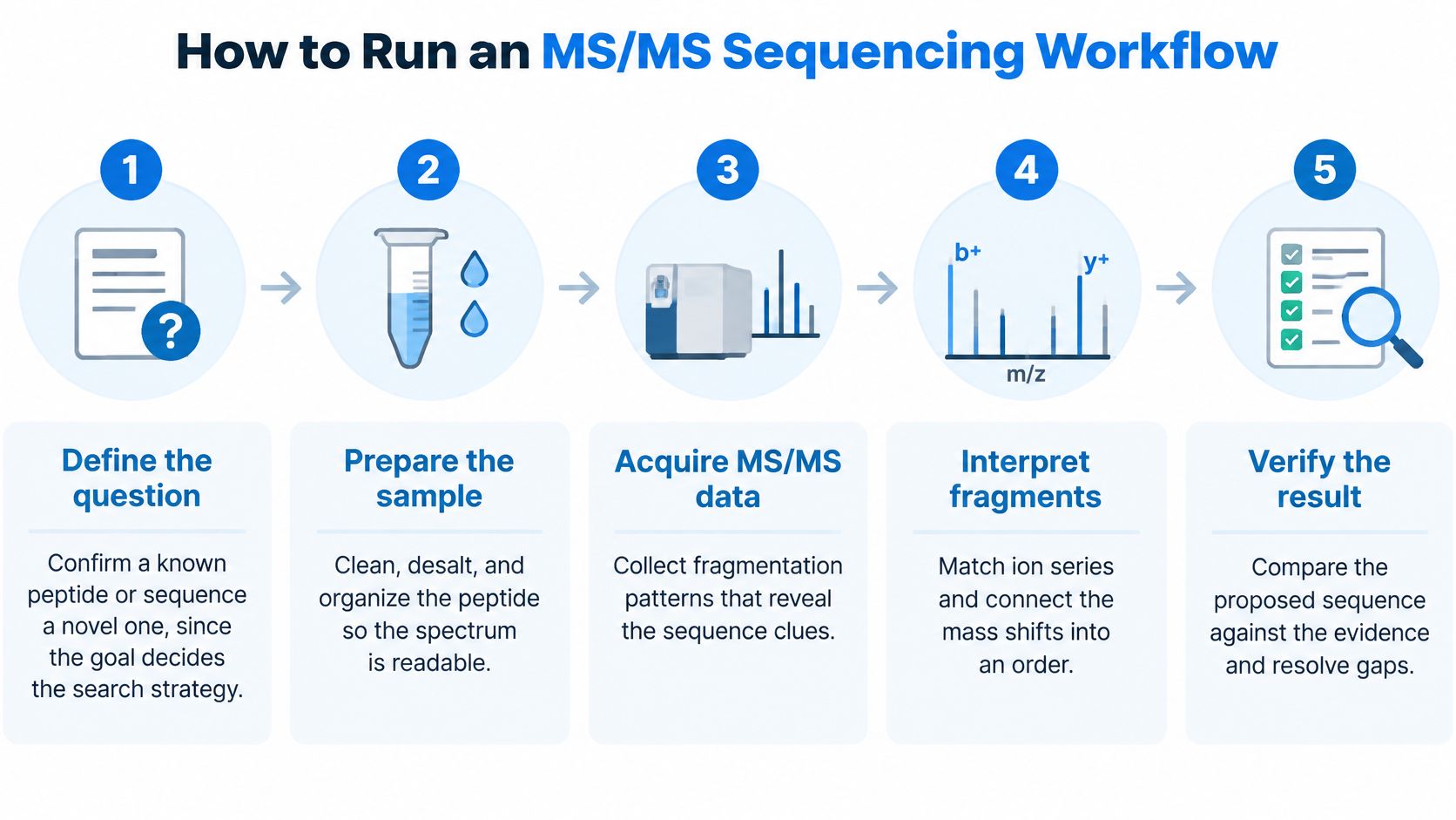
The workflow diagram summarizes the main stages; the operational record should contain the details behind each stage.
Acquisition and interpretation steps
Define the analytical question. Confirming a specified synthetic peptide calls for testing a proposed identity, including plausible variants and modifications. Investigating an unknown peptide requires a search strategy that doesn't force the result into an expected sequence.
Prepare and document the sample. The analyst checks purity, removes incompatible matrix components, and chooses digestion when the target originates from a larger protein. ProTecPep products must only be handled by qualified laboratory personnel familiar with appropriate research procedures and safety practices.
Separate the sample by liquid chromatography. Separation reduces the chance that unrelated peptides enter the same precursor-isolation window. It doesn't eliminate co-isolation, so precursor isolation and chromatographic peak shape still require review.
Acquire precursor and fragment spectra. MS1 measurements characterize candidate precursor ions, while MS2 measurements record their fragmentation products. The acquisition record should retain charge assignments, isolation settings, fragmentation conditions, and relevant instrument-calibration information.
Search the appropriate sequence space. Database searching tests represented candidates, while de novo analysis proposes residue order from spectral evidence. The analyst records precursor and fragment mass tolerances, enzyme specificity, permitted modifications, and software settings so the interpretation can be reproduced.
Inspect the proposed assignments manually. Review should follow consecutive b- and y-ion series through each region rather than count scattered matching peaks. Unsupported transitions, competing assignments, and substantial unexplained signals belong in the review notes.
Produce an evidence-bearing result. The report should state fragment coverage, the confidence classification, and unresolved positions. It should also identify which follow-up acquisition or independent measurement could change the conclusion.
Preserve an auditable output
A useful coverage map aligns the proposed residues with the cleavage positions supported by assigned fragments. An unobserved position should remain visibly unsupported rather than appearing equivalent to a well-supported region.
The deliverable is a sequence claim with a coverage map and open questions, not a screenshot of the highest software score.
For example, a terminal region may be well supported while an internal segment remains compatible with alternative orders. The report should preserve those alternatives and specify whether additional fragmentation, a complementary digest, or another method is needed.
The accompanying video provides additional context for peptide-analysis workflows.
Where Edman Degradation Still Fits
Edman degradation is often framed as the older method, yet it answers a narrower and sometimes cleaner question than an MS/MS experiment: which residues occur sequentially from an accessible N-terminus? That scope makes it useful as an independent check when a mass spectrum supports only part of a chain or leaves the terminal order unresolved.
Pehr Edman introduced the method in 1950, and the Edman degradation overview documents the chemistry along with a practical limit: modern instruments can achieve high per-cycle efficiency, yet routine reads often stop around 30 residues as losses accumulate. The method reads a chain the way a proofreader reads a word by tearing off one letter at a time from the front and identifying each removed letter. High efficiency in one cycle does not remove the cumulative loss across many cycles.
How the terminal readout works
The chemistry starts by labeling the free N-terminal amino group. Cleavage releases the terminal residue, which is identified through its phenylthiohydantoin derivative. Repeating the reaction advances the readout along the peptide.
The identity of each released derivative and its position in the reaction sequence provide the ordering information. The readout is chemical evidence for residue order, not a direct photograph of the molecule. Unlike a database match, it does not require the complete expected sequence to be supplied beforehand. That independence is the reason to keep Edman in the method set, even when MS/MS carries the main sequencing work.
Conditions that determine success
Terminal accessibility comes first. Acetylation, pyroglutamate formation, or another blocking group can prevent labeling. A failed readout is therefore inconclusive: it does not prove the sample contains no peptide.
Sample purity matters just as much. A mixture can release different residues at the same reaction stage, making the signals hard to assign to one chain. A chromatographically clean fraction gives a more interpretable trace than an unresolved preparation.
Disulfide-rich samples generally require reduction and often alkylation before analysis. Document those steps. Determining a reduced chain's sequence is not the same as establishing the original disulfide connectivity.
Method-selection insight: An independent terminal readout can resolve a specific question without establishing the complete molecule.
For a purified short peptide with an accessible terminus, Edman analysis can corroborate an N-terminal assignment or reveal unexpected processing. MS/MS is usually the better choice for mixtures, modified molecules, and broader sequence characterization, but its coverage still has to be demonstrated. Choose between them by asking which disputed position the evidence must settle, not by assuming the newer instrument always produces stronger proof.
Judging Confidence and Confirming the Sequence
An identification isn't a verified sequence. It also doesn't establish purity, correct folding, potency, or the complete modification state. A top-ranked candidate means that a search procedure preferred that candidate under specified assumptions, not that every residue has been independently demonstrated.
A 2025 IJCAI study on de novo peptide sequencing found that many errors were associated with limited fragment-ion coverage and were largely independent of the model used. Changing software may change the proposed answer without supplying the missing experimental evidence.
Separate coverage from confirmation
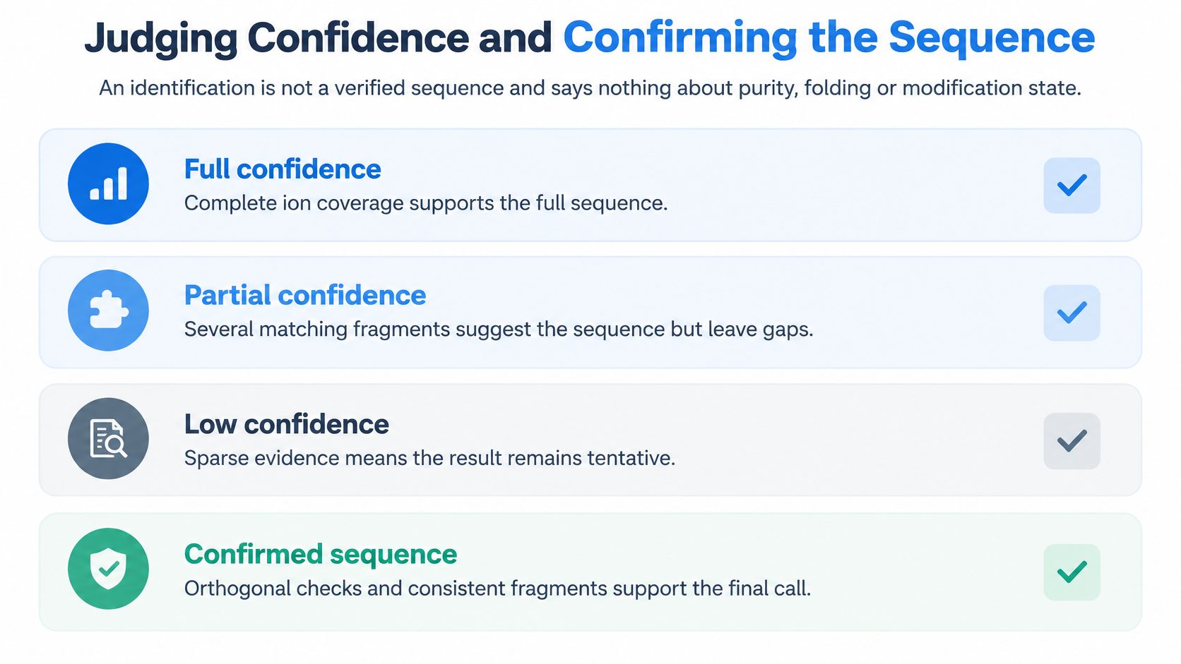
A practical confidence scheme distinguishes the extent of sequence support from the availability of independent confirmation:
- Full sequence support: Complete interpretable ion coverage supports the proposed order, with residue ambiguities and modifications explicitly resolved.
- Partial sequence tag: A continuous region is supported, but the remaining order hasn't been established.
- Ambiguous region: Sparse or conflicting evidence permits competing assignments that require more measurements.
Confirmation is an additional status, not a higher software score. A partial assignment can be independently corroborated without becoming a complete sequence.
The diagram separates evidence strength from the additional work needed for a confirmed assignment.
Diagnose the weakness before repeating the experiment
Manual review should test whether consecutive fragments support the proposed transitions, whether mass errors are consistent with the instrument method, and whether major unexplained peaks suggest another species. An analyst shouldn't count every matching peak as independent evidence.
Spectral symptoms, likely causes and first checks
| Spectral symptom | Likely cause | First check |
|---|---|---|
| Few y-ions above noise | Low fragment yield or unsuitable collision energy | Re-acquire with adjusted collision energy on a cleaner sample |
| Gap of two or more residues in the ladder | Missing fragment ions | Check consecutive series from both termini and try alternative fragmentation |
| Two sequences with near-equal scores | Isobaric I/L or near-identical K/Q assignment | Seek side-chain-specific evidence and review high-accuracy masses manually |
| Mass shift not in the search settings | Unlisted modification or adduct | Run a modification search and check the shift against known chemistry |
| Top-ranked sequence with no second method | Overreliance on one software output | Confirm the disputed region with suitable Edman analysis or an authentic standard |
Database workflows also need appropriate false-discovery controls. A dataset-level false-discovery estimate doesn't prove that a particular ambiguous residue is correct, and a high local score doesn't replace control of false identifications across the search.
Choose confirmation that addresses the disputed claim
With an authentic reference standard, comparison should examine retention behavior and fragment patterns under matched analytical conditions. Matching intact mass alone is insufficient when competing sequences or modification arrangements remain plausible.
Without a suitable standard, the analyst should name an independent method and its scope. Edman sequencing can corroborate an accessible N-terminal region; a complementary digest can provide overlapping sequence evidence; targeted fragmentation can address a poorly supported internal segment. None should be described as resolving more than the measurement covers.
A confirmed residue order still leaves other quality attributes to investigate. Peptide purity testing addresses sample composition, while folding, connectivity, and modification placement require appropriately designed characterization.
Permitted research with ProTecPep includes analytical testing, chemical characterization, method development, reference-standard comparison, stability testing, instrument calibration, in-vitro laboratory investigation, and other lawful non-clinical research activities.
Report the claim before comparing it with expectations
The report should state the observed conclusion before discussing agreement with a published sequence. That ordering helps prevent an expected identity from concealing unsupported residues.
A reusable reporting record includes:
- Method: Instrument method, search approach, and relevant interpretation settings.
- Sample state: Intact, digested, reduced, alkylated, or otherwise modified during preparation.
- Coverage percentage: The value and its definition, distinguishing supported cleavage positions from identified residues.
- Confidence level: Full support, partial tag, or ambiguous assignment, with the reason.
- Confirmation used: Reference-standard comparison or a named independent method and its scope.
- Unresolved residues: Alternative identities, uncertain positions, and proposed follow-up measurements.
A defensible sequence statement makes uncertainty visible. It allows another researcher to distinguish what the laboratory established from what remains a working hypothesis.
ProTecPep supplies research compounds for laboratory investigation, with independent third-party analytical testing and Certificates of Analysis where available. Qualified researchers can visit Pro Tec Pep to review available materials and documentation when planning sequence-analysis or chemical-characterization work.