Hydrolysis of Peptide Bonds: Mechanisms and Rates
Learn hydrolysis of peptide bonds, including acid, base, and enzymatic mechanisms, kinetics, experimental conditions, and how labs measure and limit
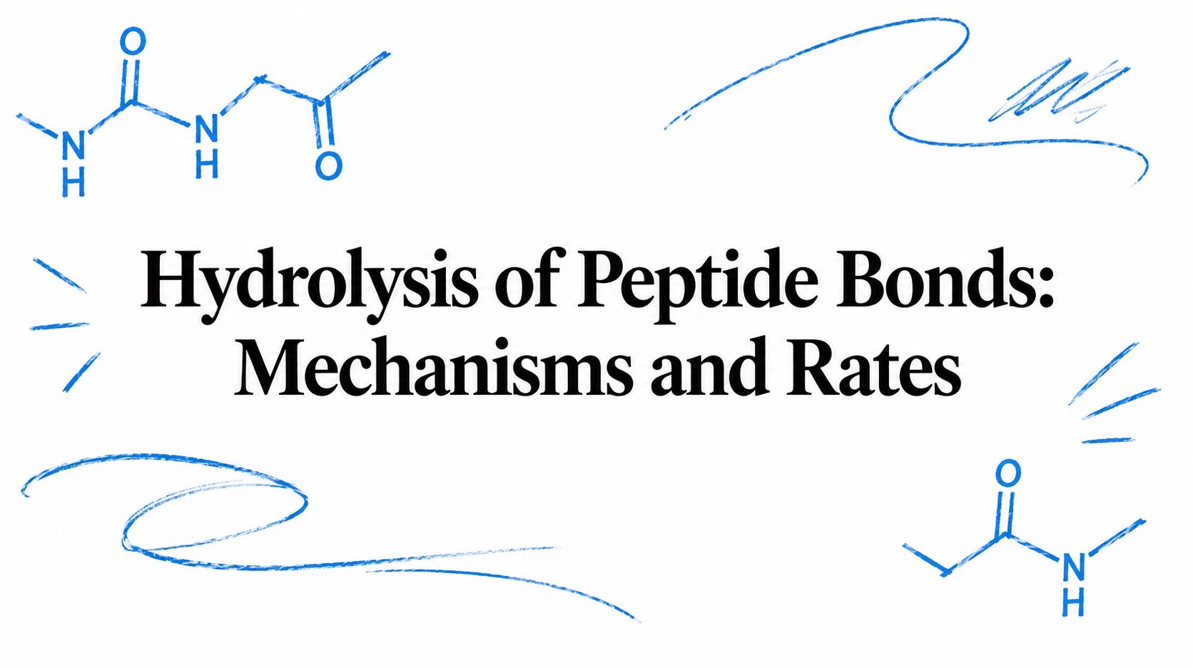
A peptide bond can sit in neutral water for centuries. A classic kinetic study estimated half-lives of about 500 years for the C-terminal bond of acetylglycylglycine, 600 years for an internal peptide bond in acetylglycylglycine N-methylamide, and 350 years for glycylglycine at 25 °C (JACS kinetic study). That single fact explains why peptide-bond hydrolysis is a chemistry problem, a sample-handling problem, and an assay-design problem at the same time.
What Peptide Bond Hydrolysis Means
Peptide-bond hydrolysis is the reaction in which water cleaves the amide linkage joining two amino-acid residues. At the bond level, one C-N bond and one O-H bond break, while one C-O bond and one N-H bond form. Atoms from water therefore become part of the products (mechanistic overview). Dissolution only disperses molecules. Hydrolysis changes the covalent backbone.
Why the bond resists water
Two features slow the uncatalyzed reaction. Water is a weak nucleophile, and resonance stabilization in the amide linkage gives the carbonyl carbon less chemical incentive to react (JACS kinetic study). The reported activation energy for uncatalyzed hydrolysis is about 96 to 105 kJ/mol (mechanistic overview). That high barrier explains why an intact peptide can remain in aqueous solution without quickly splitting.
A plain neutral buffer therefore favors preservation of the sample, not rapid self-cleavage. Hydrolysis still proceeds, but slowly enough that a visually unchanged tube may contain gradual backbone degradation.
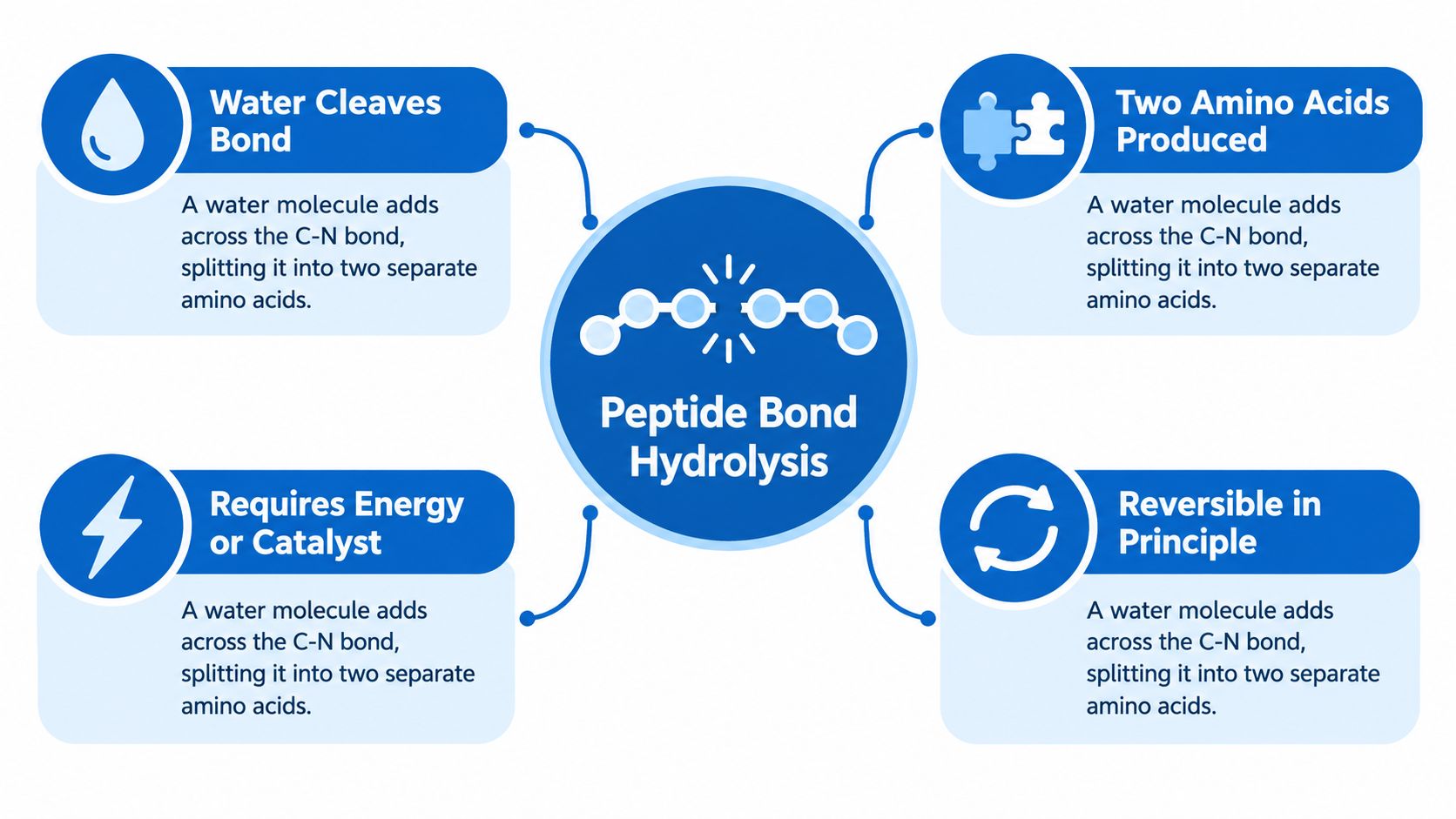
That distinction guides lab decisions. Researchers needing cleavage on a practical timescale use proteases or chemically forcing conditions, then account for the resulting handling risks. Researchers preserving a peptide instead control exposure to conditions that lower the barrier, because temperature, pH, and reactive catalysts can turn a slow background reaction into measurable degradation.
Acid and Base Catalyzed Hydrolysis
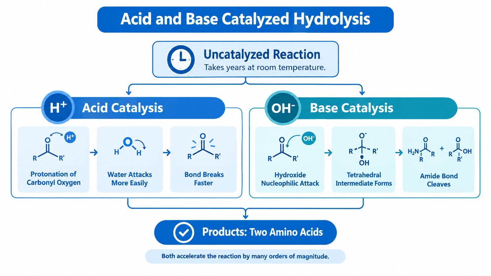
Acid and base catalysis accelerate peptide-bond hydrolysis by changing the attacking species and stabilizing the transition state. In water, both proton and hydroxide activity can influence the reaction, a behavior described by the bicatalytic model. The laboratory consequence is practical: changing pH can move a sample from slow background degradation to deliberate cleavage.
What acid catalysis does
Acid catalysis commonly begins when a proton attaches to the carbonyl oxygen or amide nitrogen. The carbonyl then becomes more electrophilic, allowing water to attack more readily. Proton transfers help the tetrahedral intermediate reorganize and release the amine-containing product, while water supplies the oxygen incorporated into the hydrolysis products. The peptide bond is unchanged in its final chemistry, but its activation barrier is lower.
What base catalysis does
Under alkaline conditions, hydroxide acts as a stronger nucleophile than neutral water. It attacks the peptide carbonyl, forming a tetrahedral intermediate that can collapse toward cleavage products. The same route can increase unwanted reactions or alter sensitive residues, so alkaline treatment requires control of exposure time, temperature, and sample composition. Acid and base hydrolysis therefore serve as forcing conditions, not preservation conditions.
| Representative rate context | Condition | Rate constant (s^-1) | Half-life | Source |
|---|---|---|---|---|
| Spontaneous cleavage | Neutral water | as low as 10^-11 (JACS kinetic study) | Centuries, depending on bond and sequence | Uncatalyzed peptide hydrolysis |
| Enzymatic cleavage | Zinc protease activity | near 10^4 (zinc-enzyme catalysis/06%3A_Enzymes/6.07%3A_Metalloenzymes)) | Extremely short on lab timescales | Proteolytic catalysis |
The contrast determines handling. A neutral aqueous sample may tolerate routine manipulation because spontaneous cleavage is slow, whereas forced acid or base treatment demands a defined endpoint and prompt neutralization or analysis. The Pro Tec Pep's research catalog provides broader context for peptide-research workflows in which stability affects compound handling.
How Proteases Cleave the Same Bond Differently
Proteases can break the same peptide bond through different chemical routes. The catalytic class determines which atoms move, how the activation barrier is lowered, and how quickly the substrate's effective half-life falls during digestion. Serine, cysteine, and threonine proteases use a nucleophilic group from the enzyme, whereas metalloproteases and aspartic proteases activate water directly.
The serine-protease pathway
A serine protease first uses a histidine residue as a general base. Histidine removes a proton from the serine hydroxyl, allowing oxygen to attack the substrate carbonyl. The tetrahedral intermediate then collapses, releasing one product and leaving an acyl-enzyme intermediate. A second, activated water molecule attacks that intermediate, releasing the remaining product and restoring the enzyme (protease-class mechanism).
This two-stage route lowers the effective activation barrier at the active site. Under suitable buffer and temperature conditions, the targeted bond's working half-life can become short, so samples may require controlled enzyme exposure and prompt quenching.
The water-activation route
Metalloproteases and aspartic proteases position water beside the scissile bond and make it a stronger nucleophile. They do not rely on the same covalent acyl-enzyme intermediate. Metal coordination or acidic residues help arrange proton transfers, so the activation barrier and useful half-life depend on the metal, pH, substrate, and inhibitors present.
That difference changes bench decisions. A metal-dependent enzyme may lose activity after chelator addition, while a cysteine protease may require a suitable redox state. Swapping trypsin for chymotrypsin therefore changes cleavage sites, not merely digestion speed. Choose the enzyme for its substrate preference and mechanism before adjusting incubation time.
Kinetics That Decide Whether a Bond Lasts or Breaks
A pH profile often reveals more about cleavage risk than a single incubation time. Reported kcat/Km profiles for proteases can be bell-shaped, with an acidic apparent pKa near 6 and an alkaline limb near 9 (zinc-enzyme comparison/02:_The_Reaction_Pathways_of_Zinc_Enzymes_and_Related_Biological_Catalysts/2.11:_Peptide_Hydrolysis)). The rate therefore depends on which catalytic groups are protonated, as well as on how the active site positions the peptide. A buffer change can alter cleavage even when the substrate and temperature stay constant.
Activation energy adds a second practical guide. For sequence-specific routes such as aspartyl peptide-bond hydrolysis, reported values are about 15 to 20 kcal/mol for model peptides and about 23 kcal/mol for monoclonal-antibody systems (sequence-dependent cleavage study). A higher barrier generally means greater temperature sensitivity, so accelerated-stability studies often vary both pH and temperature. A bond that survives a short bench exposure may still become a measurable degradation site during warm storage.
The handling consequence depends on the route. Uncatalyzed hydrolysis in neutral water is slow enough that ordinary sample preparation may preserve the backbone, whereas enzyme-mediated cleavage can occur on digestion timescales. For a protease assay, record the buffer, temperature, enzyme-to-substrate ratio, and quench step. Those details determine whether a measured fragment reflects the intended reaction or uncontrolled time in the tube. Reference information on peptide behavior can also support decisions in a compound research library.
| Condition type | What the number tells a researcher |
|---|---|
| Uncatalyzed hydrolysis | The bond can remain intact for a long time in neutral water |
| Enzymatic hydrolysis | Catalysis can make cleavage rapid on lab timescales |
| Bell-shaped pH response | Protonation state controls part of the rate |
| Activation-energy differences | Sequence context changes temperature sensitivity |
A rate constant is therefore a planning tool. Use it to set digestion timing, protect untreated samples, and distinguish chemical degradation from handling time.
Choosing Hydrolysis Conditions for Valid Amino Acid Analysis
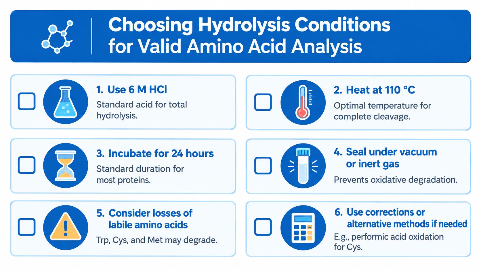
A 6 N HCl, 24 h, 110 °C run can destroy tryptophan entirely while leaving alanine recovery near 100%. That example shows why a hydrolysis result is not automatically a faithful amino-acid profile. Standard acid hydrolysis breaks peptide backbones efficiently, but concentrated acid and heat can also modify or destroy labile residues. The recovered composition may therefore differ from the starting peptide, as described in this clinical chemistry guidance.
Match the chemistry to the sequence
Start with the peptide sequence, then choose the treatment. A sequence containing labile residues may need conditions that preserve those residues, while a more stable backbone may tolerate stronger or longer hydrolysis. Bond environment, neighboring residues, temperature history, and sample matrix all affect the products collected from the tube. One universal condition rarely gives a complete answer.
The activation energy and expected half-life also guide the handling decision. A route with a lower barrier may require rapid quenching and cold storage after cleavage. A slower route may preserve the sample during preparation but still cause selective loss during prolonged heating. Pair the chosen conditions with the expected residue stability rather than treating hydrolysis as total backbone removal.
Validate recovery instead of assuming it
Include sequence-matched recovery controls, freeze aliquots of standards, and confirm the result with an orthogonal readout such as intact-mass analysis. A high apparent recovery can conceal selective loss, distorting purity or net-content interpretation. The practical question is whether the assay measures the complete peptide composition or only the residues that survived treatment.
Enzymatic digestion may preserve some acid-labile residues, yet it can leave partial cleavage when the substrate is folded or the local bond context resists attack. Choose the route that fits both the sequence and the measurement, then document time, temperature, reagent strength, and quench conditions so the result remains interpretable.
Demasking: Why Substrate Access Controls Digestion Speed
A slow digestion can begin with substrate access. Quantitative proteolysis work separates a non-hydrolytic demasking step, in which a protein opens enough to expose the target bond, from the chemical cleavage that follows (demasking model). A folded substrate, aggregate, or membrane-associated target can therefore delay digestion even while the protease remains active.
Access creates the lag phase
Protease activity alone does not guarantee rapid cleavage. If the substrate stays folded or buried, the enzyme waits for a workable binding position, much like a tool held against a covered surface. Time-course sampling can reveal this delay, whereas a single endpoint cannot show whether cleavage began late or proceeded slowly throughout.
Pair intact-mass measurements with fragment analysis when the distinction matters. The two readouts help separate limited access from resistance at the peptide bond itself.
Sequence context redirects cleavage
Aspartyl cleavage shows how local chemistry can change both rate and sample handling. In one documented pathway, the ionized side-chain carboxylate of an aspartate residue attacks a neighboring peptide bond and promotes backbone fragmentation (sequence-dependent cleavage study). Under the study conditions, hydrolysis at pH 1 was about twice as fast as at pH 3. A small pH change can therefore alter the fragments that reach the chromatogram.
Record substrate conformation, pH, temperature, enzyme-to-substrate ratio, mixing, and sampling time. These details connect the mechanism to the bench decision: improve exposure, adjust the chemical environment, or shorten the incubation before secondary cleavage changes the sample.
Storage and Handling to Reduce Degradation
Storage conditions determine which cleavage routes remain active. Lyophilized storage at -20 °C slows reactions with activation barriers, while aliquoting prevents repeated freeze-thaw cycles from repeatedly warming the sample. Prompt reconstitution in a compatible solvent, cold-chain shipping, and rapid processing protect the peptide before analysis begins.
Match the solvent to the sequence. Aqueous buffers generally suit hydrophilic peptides, whereas dilute organic co-solvents can improve handling of hydrophobic sequences. Keep pH near neutrality when the aim is to suppress acid- and base-catalyzed hydrolysis. For solvent and storage specifications on individual compounds, the research peptide catalog lists solubility and recommended storage for each product.
Handle storage as chemistry, not logistics
Every handling choice has a chemical consequence. Cold temperature slows bond cleavage, low-water formulations limit hydrolysis, and aliquoting reduces exposure to warming and refreezing. A useful bench rule is simple: keep the peptide dry and cold until use, then minimize the time it spends reconstituted before measurement.
Forced Degradation Studies and What They Tell You
Forced degradation studies turn the mechanism into a map. By exposing a peptide to high temperature, acidic or alkaline pH, oxidative stress, or metal exposure, researchers can see which cleavage products appear first and infer the dominant vulnerability of that sequence. The testing resource at Pro Tec Pep's testing overview fits naturally with that workflow because the question is always the same, what exactly changed in the lot that arrived at the bench?
Read the products, not just the starting material
An observed aspartyl cleavage points toward a sequence motif that's chemically primed to attack itself under the wrong conditions. A diketopiperazine signal points toward a different kind of intramolecular rearrangement, not just random backbone loss. Those signatures matter because they tell the researcher whether the risk is sequence-based, pH-based, or tied to a specific storage event.
Tie the result back to the workflow
The main lesson is simple. Pick hydrolysis conditions by sequence, control storage conditions by mechanism, and verify completeness with orthogonal readouts instead of assuming that one endpoint measurement proves the whole sample behaved well. Purity and stability are lot-specific properties, not abstract promises, so the only defensible interpretation is the one linked to the actual handling history.
Pro Tec Pep supports research teams that need batch-linked documentation, cold-chain handling options, and analytical transparency for peptide and related compounds. For laboratories working through hydrolysis, digestion, or stability questions, that kind of documentation helps connect the chemistry at the bench to the lot that was received. Visit Pro Tec Pep to review research-use offerings and supporting product information.