What Is IGF-1 LR3 Structure Mechanism and Research Use
What is IGF-1 LR3? Learn how its 83-residue structure, receptor mechanism and research applications differ from native IGF-1 in this lab-focused guide.

IGF-1 LR3 is an 83-residue engineered analog of native human IGF-1, which has 70 residues, with an Arg substitution at position 3 and a 13-amino-acid N-terminal extension that reduces IGFBP binding and extends persistence. In laboratory research, it's studied as a recombinant growth-factor analog for sustained IGF-1 receptor signaling, not as an approved clinical therapy.
Why would researchers redesign a molecule that already activates the IGF-1 receptor? The answer is that native IGF-1 can be difficult to study when binding proteins sequester much of the ligand and its circulation time is short. IGF-1 LR3 was engineered to change that experimental problem at the molecular level. The resulting reagent can provide a longer signaling window, but longer persistence still doesn't guarantee a useful outcome in a complex biological model.
Introduction to IGF-1 LR3 for Laboratory Research
What changes when a researcher needs IGF-1 receptor signaling to remain measurable for longer than native IGF-1 allows? Native IGF-1 interacts with IGF-binding proteins, including IGFBP-3, and is often described as having a circulation time of roughly 10 minutes. Sustained receptor stimulation can therefore require repeated exposure or continuous infusion. A review of IGF-1 LR3 describes the engineered analog as having a substantially longer half-life and about a 3-fold increase in potency compared with native IGF-1 (peer-reviewed review of IGF-1 LR3 structure and function).
The design can be understood as changing the ligand's handling rather than adding more native IGF-1. An arginine substitution at position 3 and a 13-amino-acid extension at the N-terminus produce an 83-residue peptide, compared with the 70-residue native IGF-1 molecule. These modifications reduce interactions with IGF-binding proteins and alter how much ligand remains available for receptor interaction. The sequence details are documented in a technical product datasheet (engineered IGF-1 LR3 sequence datasheet).
Research boundary: A longer signaling window is a pharmacological property, not proof of clinical benefit.
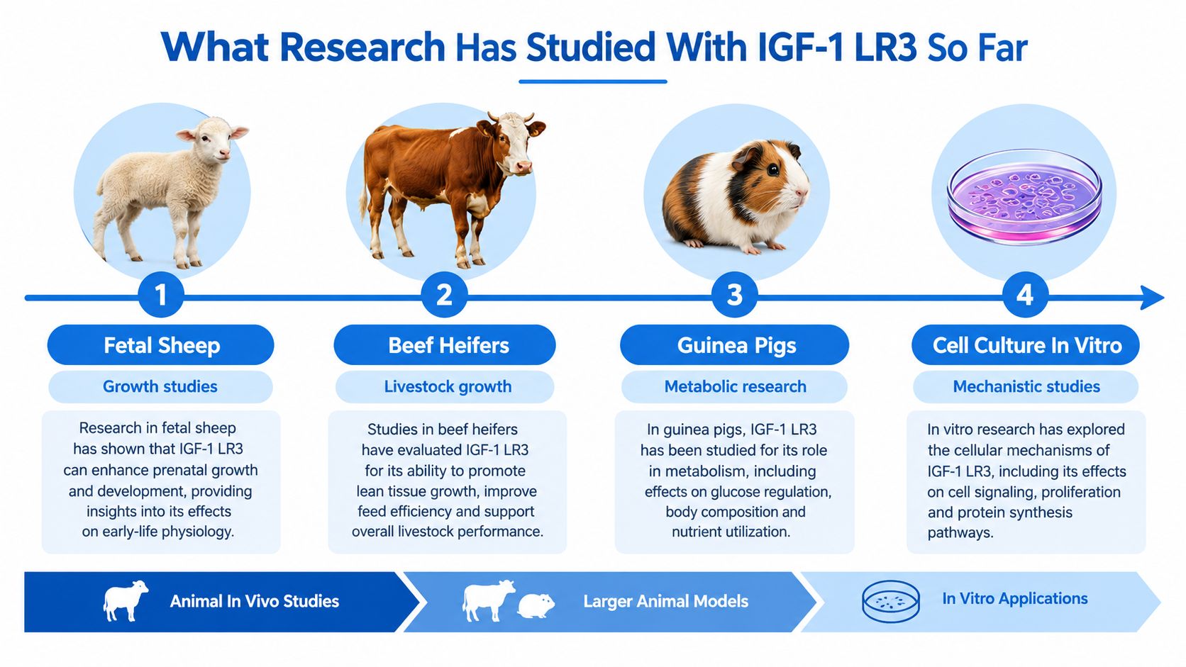
IGF-1 LR3 is therefore best treated as a laboratory reagent. Published work has examined it in animal models and cell-based systems involving growth, metabolism, and receptor signaling. Reported models include fetal sheep, beef heifers, guinea pigs, neonatal pigs, and in-vitro systems. Those models help define biological responses, but they do not establish a safe or effective human application (historical and research overview of IGF-1 LR3).
A sound interpretation follows the chain from structure, to binding behavior, to receptor signaling, and then to the limits of each experimental model. The same logic applies to certificate-of-analysis specifications: they help researchers judge identity, purity, and suitability for a protocol, not predict personal outcomes.
How IGF-1 LR3 Differs From Native IGF-1 in Structure
What changes when a peptide keeps the receptor-binding core of IGF-1 but gains a redesigned outer structure? IGF-1 LR3 answers that question through two sequence modifications that distinguish it from native human IGF-1.
Native IGF-1 contains 70 amino-acid residues. IGF-1 LR3 contains 83 residues, including a 13-amino-acid N-terminal extension. “Long” refers to this added segment, while “R3” identifies an arginine substitution at position 3.
A key analogy is a key with a modified handle. The teeth still represent the features involved in receptor interaction, while the redesigned handle changes how other proteins can bind, transport, or release the molecule. IGF-1 LR3 is therefore a related ligand with altered pharmacology, not native IGF-1 at a higher concentration.
The two defining modifications
The first modification is a Glu3 to Arg substitution, written as Glu3→Arg or E3R. The second is the 13-residue N-terminal extension. Technical characterization describes these changes while retaining the core receptor-binding region (recombinant IGF-1 LR3 datasheet).
The sequence differences can be summarized clearly:
- Residue count: Native IGF-1 has 70 residues; IGF-1 LR3 has 83.
- Position 3: IGF-1 LR3 contains arginine where native IGF-1 contains glutamic acid.
- N-terminal region: The analog carries a 13-amino-acid extension.
- Production context: The peptide is produced in recombinant systems for controlled laboratory investigation.
- Reported mass: Technical descriptions place its molecular mass at about 9.1 kDa.
Why the extension matters
The added residues and position-3 substitution change more than the peptide's length. They reduce affinity for IGF-binding proteins, particularly IGFBP-3, shifting the balance between protein-bound and receptor-available ligand. The receptor-binding core remains relevant, while the surrounding design affects how readily the peptide is sequestered.
Researchers should read the sequence as a functional design rather than only an identification label. Sequence, molecular mass, formulation state, and lot-specific analytical results help determine whether a material matches the intended research reagent.
That distinction matters when comparing experimental results. Although native IGF-1 and IGF-1 LR3 can engage the IGF-1 receptor, their different binding-protein interactions may alter exposure, assay timing, concentration-response behavior, and interpretation of downstream readouts. Structural similarity therefore does not make the two ligands interchangeable.
Mechanism of Action and Why Binding Protein Evasion Matters
Could a small sequence change alter how long a growth factor remains available to a receptor? IGF-1 LR3 illustrates that structure-to-function relationship through a clear chain:
- Its engineered sequence has lower affinity for IGF-binding proteins.
- A larger fraction can remain free and available to receptors.
- That availability may extend receptor signaling in suitable experimental systems.
- Assays may therefore show stronger or more sustained exposure-dependent responses.
- Receptor activity still requires biological interpretation, because it does not establish a tissue-level effect.
Native IGF-1 operates within a regulated binding environment. IGF-binding proteins influence the amount of free ligand, its movement through a system, and its access to receptors. IGF-1 LR3 has reduced interactions with these proteins, particularly IGFBP-3, shifting the balance toward free ligand (technical summary of IGF-1 LR3 binding behavior).
Receptor availability versus biological outcome
The IGF-1 receptor is a cell-surface receptor connected with signaling pathways involved in growth and metabolism. When a ligand stays available to that receptor for longer, researchers can examine sustained pathway engagement without continuous infusion. That property helped establish IGF-1 LR3 as an experimental tool.
Technical descriptions place IGF-1 LR3's half-life in the 20 to 30 hour range, compared with the roughly 10-minute half-life commonly cited for native IGF-1 (review of IGF-1 LR3 pharmacology). A longer exposure profile does not resolve questions about tissue specificity, feedback regulation, metabolic effects, or functional outcomes.
The commonly cited potency difference needs the same careful framing. A peer-reviewed review describes IGF-1 LR3 as having an around 3-fold increase in potency versus native IGF-1, associating this difference with reduced affinity for IGF-binding proteins. In an in-vitro assay, that property may make sustained receptor signaling easier to observe. In an animal model, the result can still vary with developmental stage, tissue distribution, nutrient status, feedback loops, and treatment duration.
Strong receptor signaling is an observation. It isn't the same as a demonstrated therapeutic effect.
Sound experimental design separates receptor-proximal measurements, such as pathway activation, from distal outcomes, including growth, metabolism, cognition, or tissue remodeling. IGF-1 LR3 may produce a clear molecular signal while the functional result remains mixed or absent.
What Research Has Studied With IGF-1 LR3 So Far
What can IGF-1 LR3 experiments show? The answer depends on the model, the endpoint, and the exposure conditions. Early recombinant fusion-protein work established the analog's enhanced biological activity relative to native IGF-1. Later patent activity reflected commercial and application interest, but neither history nor patenting makes a research analog an approved therapy.
Published studies have examined IGF-1 LR3 in several settings:
- Fetal sheep: Growth and fetal-development studies, including growth-restriction models.
- Beef heifers: Livestock growth and metabolic physiology.
- Guinea pigs: Metabolism and growth-related biology.
- Neonatal pigs: Developmental and metabolic research.
- Cell culture: Receptor signaling, binding behavior, and controlled exposure responses.
Each model functions like a different instrument. Cell systems isolate receptor and pathway behavior with limited biological interference. Animals add circulation, endocrine feedback, organ interactions, nutrition, and developmental stage. Findings therefore require model-specific interpretation rather than direct transfer to another species or to humans.
The importance of negative and mixed findings
A recent physiology study illustrates why molecular activity should be separated from functional improvement. In a 1-week IGF-1 LR3 treatment study of growth-restricted fetal sheep, treatment did not improve fetal growth, insulin secretion, or β-cell mass. It also reduced circulating amino acids, especially branched-chain amino acids (2025 fetal growth restriction study).
The result indicates that the analog altered physiology, while the measured growth-related outcomes remained unchanged within that model and time frame. A detectable response is therefore not automatically a beneficial one.
A separate 2025 mouse study involving intranasal LR3-IGF-1 examined cortical amyloid plaque remodeling and cognitive outcomes. Its findings should be cited to the study that performed that mouse experiment, not to the fetal sheep paper. More broadly, tissue-level remodeling and behavioral preservation represent different endpoints, so one cannot substitute for the other.
The evidence remains preclinical. A research-status summary reports no human clinical trials or randomized controlled trials of exact-substance IGF-1 LR3, with the available literature centered on recombinant expression work and animal studies (summary of IGF-1 LR3 research status). This limitation should shape experimental design: COA specifications can help define material identity and assay inputs, but they cannot predict personal outcomes or convert preclinical findings into clinical evidence.
Stability Handling and Laboratory Sample Preparation
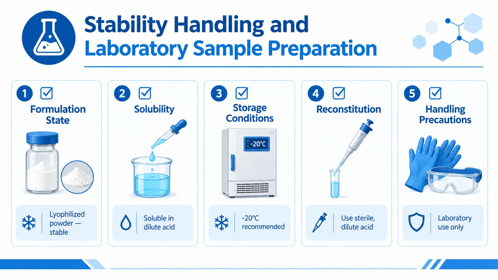
How should a laboratory treat IGF-1 LR3 once it leaves the vial? The answer depends on whether the material is still a lyophilized powder or has entered an aqueous solution. That distinction works like separating a dry reagent from a prepared assay mixture: each state has different handling risks and documentation needs.
Before preparation, record the vial's lot, confirm that it is sealed and protected from light, and inspect it for visible damage. Link these observations to the batch Certificate of Analysis. A practical workflow then separates dry-material storage from solution handling:
- Formulation state: Record the powder's appearance and lot identifier.
- Solubility: Technical handling summaries describe dilute acid, such as dilute acetic acid, as a suitable solvent environment for laboratory dissolution.
- Addition method: Add solvent slowly along the vial wall. Use gentle swirling rather than vigorous shaking.
- Solution inspection: Check the solution for clarity and visible particulates before adding it to an assay.
- Aliquot planning: Use aliquots to reduce repeated warming and freeze-thaw exposure.
- Buffer compatibility: If an acidic stock enters an assay buffer, include its time in dilute neutral conditions in the stability plan.
Storage logic
Product documentation and laboratory handling summaries commonly recommend -20°C storage for dry material. Keep the vial sealed and protect it from moisture and light. Reconstitution changes the control problem: the peptide is now in an aqueous environment, where time, temperature, container contact, and buffer composition may affect the sample.
Sample-preparation rule: Put a storage condition in the protocol only when it is linked to formulation state, preparation time, and the assay's validated stability window.
Use lot documentation or validated supplier handling data for the stability timeframe. Avoid treating a generic storage statement as proof that every prepared solution remains suitable for every assay. Record the preparation date, solvent, concentration, container type, storage temperature, aliquot identifier, and freeze-thaw history.
These records make interpretation more disciplined. If an assay result shifts, the team can examine biological variation alongside degradation, aggregation, adsorption to the container, or altered buffer compatibility. The controls described here apply to in-vitro laboratory sample preparation only. They are not instructions for administration to humans or animals.
How Batch COAs and Purity Specs Inform Experimental Design
A Certificate of Analysis is part of experimental control, not only a purchasing record. For IGF-1 LR3, a batch-specific COA links the material to its identity, composition, purity, and stated handling requirements. Verify the exact lot instead of relying on a generic product page.
Review the document before the experiment begins:
| Documentation Element | What to Verify | Design Implication |
|---|---|---|
| Identity | Peptide name, sequence, CAS, and analytical identity result | Confirms that the reagent matches the intended IGF-1 LR3 analog |
| Purity | HPLC method, reported purity, and chromatogram where available | Helps define impurity limits and interpret assay variability |
| Net content | Declared peptide content for the specific lot | Supports concentration normalization and material accounting |
| Sterility | Whether sterility testing applies to the supplied research material | Prevents unsupported assumptions about microbial status |
| Endotoxins | Lot-specific endotoxin result or stated testing status | Matters for cell-based and sensitive biological assays |
| Heavy metals | Reported heavy-metal testing and result | Helps identify non-peptide contaminants that could affect readouts |
| Conformity | Agreement between product description and released batch data | Supports procurement records, method reporting, and audit trails |
Reading purity in context
A declared threshold such as ≥98% describes a specification. It does not mean that every assay response will be identical. Identify the analytical method, determine whether the value represents chromatographic purity or another measure, and record the lot-specific result used in the study design.
Purity also needs to be separated from identity. A sample can meet a reported purity criterion while still requiring confirmation that its sequence and molecular mass match the intended analog. Technical documentation places IGF-1 LR3 near 9.1 kDa in mass, while a lot record may report a more detailed molecular-weight value and provide mass-spectrometric identity confirmation. Preserve those details in the experimental record rather than listing only the product name. The IGF-1 LR3 technical datasheet can serve as a reference for the type of technical information to review.
COA values guide experimental design in practical ways. Net content affects concentration calculations and material accounting. Endotoxin and sterility information determines whether the supplied material is suitable for the planned cell system and which controls are needed. Conformity records connect the released lot to the methods section and later audit review.
Before preparing the first plate, ask:
- Does the sequence match the intended analog?
- Does the formulation state fit the planned sample-preparation method?
- Are purity and net-content values adequate for the assay?
- Are sterility and endotoxin results relevant to the cell system?
- Can the lot be traced in the final methods section?
Used this way, the COA becomes part of the evidence chain. It helps distinguish a biological difference from a material-quality difference and makes the experimental result easier to interpret.
Common Misconceptions and Research Use Only Boundaries
Could a longer signaling window be mistaken for a better research result? Longer persistence does not automatically predict improved outcomes. IGF-1 LR3 is described as having greater potency than native IGF-1, yet recent animal work found that strong biological activity did not improve growth, insulin secretion, or β-cell mass in the tested fetal sheep model. This separates receptor-level activity from a whole-organism outcome. The peer-reviewed review of structure and potency provides context for the structure and potency claims.
Half-life statements need equally careful reading. Human pharmacokinetic data for IGF-1 LR3 itself are unavailable. The often-repeated about 20-hour value has been linked to native recombinant human IGF-1, not to direct human measurement of LR3, as discussed in this review of IGF-1 LR3 pharmacology and evidence gaps. Controlled human safety data for the exact substance are also absent. Hypoglycemia, intracranial hypertension, lipohypertrophy, and acromegaloid soft-tissue changes are mainly inferred from related IGF-1 therapies or the wider pathway, rather than established as LR3-specific effects.
IGF-1 LR3 therefore belongs within in-vitro, analytical, and permitted preclinical laboratory research. Batch-specific COAs, validated assay conditions, institutional oversight, and model-specific evidence should guide study design. They help researchers test a defined material under defined conditions, instead of treating marketing language or unrelated IGF-1 products as evidence of personal outcomes.
For laboratory research use only. Not for human or veterinary use. Not a drug, food, or cosmetic. Not evaluated by the FDA. Not intended to diagnose, treat, cure, or prevent any disease.