Thymosin Alpha 1 Peptide Benefits from the Evidence
Thymosin alpha 1 peptide benefits in the literature: immune-marker shifts, mixed sepsis mortality data, and research-only handling checks.

Reported thymosin alpha 1 peptide benefits are immune-modulatory findings from specific studies, not established treatment effects, because sepsis mortality was 23.4% versus 24.1% in the large phase 3 trial while an earlier pooled analysis favored the peptide. That conflict is the central fact. A biomarker can move, a pooled estimate can look favorable, and a larger confirmatory trial can still find no survival difference.
Popular advice gets this backward. It starts with a long list of personal immune outcomes, treats every favorable laboratory signal as proof, and saves the null trial for a footnote, if it appears at all. That's not a useful way to read this literature. A more honest order puts the verdict first, puts the trial table immediately after it, and uses mechanism afterward to explain what those trials were testing. Researchers who reverse that order end up arguing from a plausible pathway to an unproven clinical claim.
The distinction matters for laboratory design. A peptide that changes dendritic-cell markers in culture has produced a measurable cellular finding. It hasn't established a treatment effect in people. Keeping those two statements separate protects protocols, presentations, and product pages from claims the data can't carry.
What Reported Benefits Actually Mean
Searches for thymosin alpha 1 peptide benefits usually expect a list of immune improvements a person might feel. The literature doesn't support that framing. Thymosin alpha 1, also written Tα1, is described as a 28-amino-acid thymic peptide studied as an immune-response modulator, not as a proven therapy. It belongs to the thymus-derived peptide family and shouldn't be confused with thymosin beta-4, a different compound with a different research history.
Three evidence buckets need separation before any conclusion makes sense.
- Cell-signaling findings: Laboratory work describes receptor activity, dendritic-cell maturation, and downstream immune signaling in controlled cell systems.
- Biomarker shifts: Disease-specific trials report changes in lymphocyte subsets and inflammatory markers, which show biological activity but don't prove a clinical outcome.
- Mortality results: Sepsis trials and pooled analyses point in different directions, so survival can't be presented as settled.
Those buckets explain the mixed verdict. The 2013 ETASS trial produced an early favorable signal in severe sepsis. A 2025 pooled sepsis analysis also favored Tα1. Then the 2025 phase 3 TESTS trial didn't confirm a mortality reduction. None of those results cancels the others on its own. Together, they show why trial size, comparator, endpoint, and uncertainty interval matter more than direction alone.
A measured immune change and a proven clinical benefit are different claims, and the literature on Tα1 supports the first far more consistently than the second.
The useful output for a laboratory audience is therefore a study map, not a use case. Readers designing or reviewing nonclinical and translational research can use the findings to choose endpoints, controls, and cautious language. Nothing in the discussion that follows is a human or veterinary instruction, and reported activities shouldn't be converted into personal outcomes.
Clinical Findings Side by Side
The table is the fastest way to see why broad claims fail. Readers should read the uncertainty interval before the headline direction, because several favorable-looking results are statistically fragile or contradicted by stronger designs.
Thymosin Alpha 1 Trial Findings by Endpoint
| Study | Design and size | Endpoint | Reported result | How to read it |
|---|---|---|---|---|
| ETASS 2013 | Multicenter RCT, 361 adults with severe sepsis | 28-day mortality | 26.0% vs 35.0%; RR 0.74 (95% CI 0.54–1.02); P 0.062, log-rank P 0.049 | Early signal; interval crosses 1.0 |
| Sepsis meta-analysis 2025 | 11 RCTs, 1,927 patients | 28-day mortality | OR 0.73 (95% CI 0.59–0.90), P=0.003; about 22.5% vs 28.5% | Pooled signal, not a single confirmatory trial |
| TESTS phase 3 2025 | Double-blind placebo RCT, 1,106 enrolled, 1,089 analyzed | 28-day mortality | 23.4% vs 24.1%; HR 0.99 (95% CI 0.77–1.27), P=0.93 | No significant mortality benefit |
| TESTS 90-day | Same phase 3 population | 90-day mortality | 31.0% vs 32.4%; HR 0.94, P=0.54 | Longer follow-up also null |
| Severe acute pancreatitis 2025 | 5 RCTs, 706 patients | CD4-positive percentage | Mean difference +4.53 points (95% CI 3.02–6.04) | Biomarker shift, not a cure claim |
| Severe acute pancreatitis ratio | Same 5-trial pool | CD4/CD8 ratio | Mean difference +0.42 (95% CI 0.26–0.58); CD8 change not significant | Subset balance changed; CD8 alone did not |
| CRP by regimen group | Same pancreatitis pool | C-reactive protein | Lower-amount regimens MD -30.12 mg/L; higher-amount regimens not significant | Don't convert this into an administration instruction |
The Sepsis Conflict
ETASS came first. The ETASS trial report enrolled 361 adults with severe sepsis, with 181 assigned to thymosin alpha 1 and 180 to control, and recorded 47 deaths versus 63 deaths within 28 days. Mortality was 26.0% versus 35.0%, an absolute difference of 9.0 percentage points. The relative risk was 0.74, but the 95% CI of 0.54 to 1.02 crossed 1.0. The non-stratified P value was 0.062 and the log-rank P value was 0.049, a borderline pattern rather than a clean confirmation.
That design also limits generalization. ETASS ran in a single-country setting, under sepsis definitions and background care from its period. A result can be historically important and still be too fragile to settle practice or product language. ETASS is best read as the signal that justified larger trials.
The pooled evidence looked stronger. The 2025 systematic review and meta-analysis combined 11 randomized controlled trials with 1,927 septic patients, including 967 in the thymosin alpha 1 groups and 960 controls. It recorded 218 deaths versus 274 deaths and calculated an odds ratio of 0.73 with a 95% CI of 0.59 to 0.90 and P=0.003. Observed mortality was approximately 22.5% versus 28.5%, a difference of about 6 percentage points. A related Frontiers in Cellular and Infection Microbiology meta-analysis reported the same odds ratio of 0.73 for 28-day mortality with a 95% CI of 0.59 to 0.90 and P=0.003.
TESTS then weakened that conclusion. The phase 3 TESTS trial in The BMJ enrolled 1,106 adults with sepsis, and 1,089 participants entered the modified intention-to-treat analysis, with 542 in the Tα1 group and 547 receiving placebo. Twenty-eight-day mortality was 23.4%, or 127 participants, versus 24.1%, or 132 participants. The hazard ratio was 0.99, with a 95% CI of 0.77 to 1.27 and P=0.93. Ninety-day mortality was 31.0% versus 32.4%, with a hazard ratio of 0.94 and P=0.54.
Interpretive rule: When a large double-blind trial finds almost identical mortality, earlier pooled results become a hypothesis about subgroups or context, not a settled survival claim.
The reasonable synthesis is narrow. Any apparent sepsis effect may depend on patient selection, disease phenotype, timing, or background care. Until a confirmatory trial identifies who benefits and why, Tα1 remains investigational rather than an established sepsis therapy.
The Pancreatitis Biomarkers
Severe acute pancreatitis provides a different kind of evidence. The 2025 meta-analysis indexed in PubMed pooled five randomized controlled trials involving 706 patients and reported a CD4-positive-cell percentage mean difference of 4.53 percentage points, with a 95% CI of 3.02 to 6.04 and P<0.00001. The CD4-positive/CD8-positive ratio mean difference was 0.42, with a 95% CI of 0.26 to 0.58 and P<0.00001, while the CD8-positive change wasn't statistically significant. Lower-amount study regimens were associated with a C-reactive-protein mean difference of -30.12 mg/L, with a 95% CI of -35.75 to -24.49 and P<0.00001, whereas higher-amount regimens didn't show a significant CRP difference.
The Frontiers in Immunology systematic review likewise reported that thymosin alpha 1 increased CD4+ cell percentages by 4.53 percentage points versus control, with a 95% CI of 3.02 to 6.04, across 5 randomized controlled trials involving 706 patients. Those are credible pooled biomarker findings. They aren't proof of treatment efficacy, and they aren't administration guidance for any person or animal.
How Thymosin Alpha 1 Signals Immune Cells
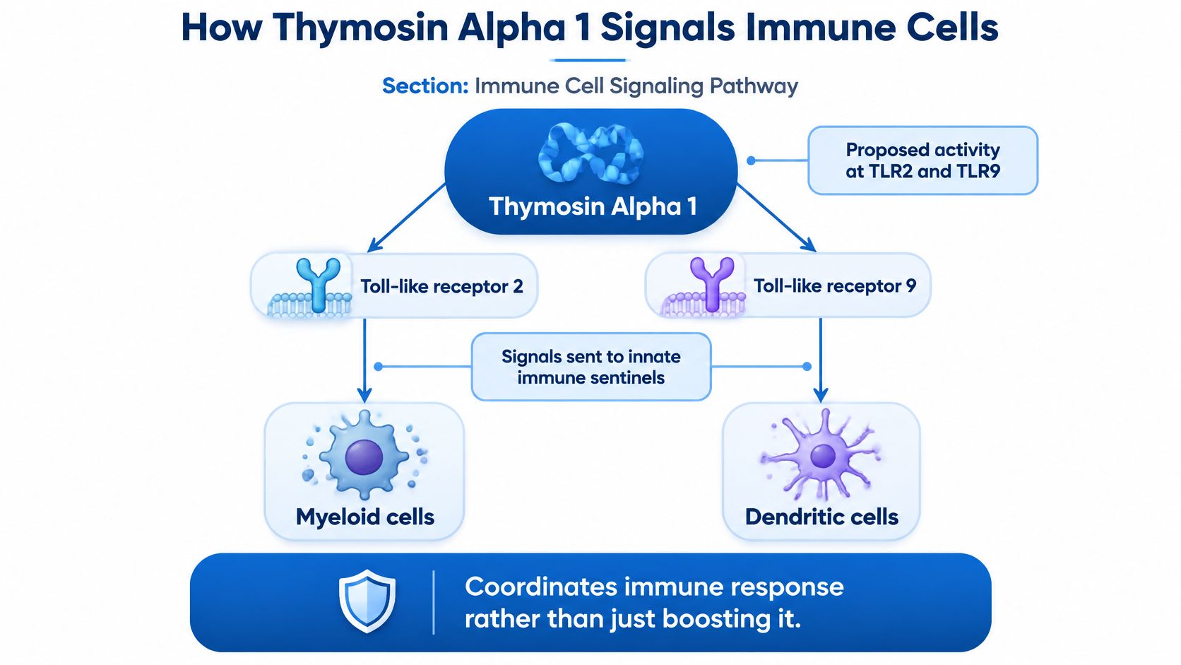
The mechanism explains why researchers keep studying Tα1 despite the conflict on mortality. It doesn't rescue a survival claim, but it does define laboratory endpoints that can be tested cleanly. The diagram below places the peptide upstream of sentinel immune cells rather than at the end of a simple stimulation pathway.
A Coordinator Model, Not a Volume Control
A nonspecific stimulant would be expected to push immune activity upward across many readouts. Tα1 is described differently. The review of thymosin alpha 1 immune activity describes activity at Toll-like receptors 2 and 9 on myeloid and dendritic cells, promoting dendritic-cell maturation, antigen presentation, and downstream cytokine signaling. Earlier clinical literature in that review reports changes in CD3+ and CD4+ cells together with altered IL-6, IL-10, and TNF-α concentrations.
That pattern fits coordination. Dendritic cells collect antigen, mature, display molecular information to T cells, and shape the next stage of the response. If a peptide changes that handoff, downstream T-cell subsets and cytokines can shift without every inflammatory marker moving in the same direction. A study that records only a total white-cell count can't detect that kind of reorganization.
Consider a typical laboratory scenario. A research team exposes dendritic cells to Tα1 under controlled in-vitro conditions and sees stronger maturation markers but little change in a broad inflammatory panel. A simplistic interpretation calls the result inconsistent. A coordinator model predicts exactly that possibility, because antigen presentation, regulatory signaling, and inflammatory output are related but not identical endpoints.
Context can also produce a flat readout. Receptor expression, baseline immune status, pathogen burden, and concomitant therapy may determine whether the peptide produces measurable restoration or limited effect. Cells with low receptor availability, an already-normal subset balance, or overwhelming background stimulation may leave little room for a separate Tα1 signal. A negative assay therefore needs the same careful interpretation as a positive one.
Endpoints That Match the Proposed Pathway
Laboratory evaluation is strongest when each endpoint corresponds to a step in the proposed sequence.
- Score dendritic-cell activation markers: Maturation markers test whether sentinel cells changed state after exposure under defined conditions.
- Record T-cell subset distribution: CD3-positive, CD4-positive, CD8-positive, and regulatory populations show whether immune balance shifted rather than merely expanded.
- Run antigen-presentation assays: Functional presentation readouts test whether marker changes translated into a different signal to T cells.
- Use multiplex cytokine panels: IL-6, IL-10, TNF-α, and related analytes can move in different directions, so a panel preserves information a single marker would hide.
The video below is useful as a visual companion to that endpoint logic, particularly for readers who separate receptor engagement from downstream cellular function.
Those endpoints also create falsifiable claims. If dendritic cells don't mature, antigen presentation doesn't change, and cytokine patterns stay flat, a general statement about immune coordination hasn't been supported in that system. If maturation rises while presentation stays flat, the pathway is interrupted downstream. That level of resolution is more valuable than a generic immune-support label.
The 2006 Dendritic-Cell Chain
The mechanistic case becomes clearer in a defined cell experiment. The 2006 mechanistic study indexed in PubMed used human and murine dendritic cells and showed that Tα1 modulated immune signaling through Toll-like receptor 9, or TLR9. TLR9 is a pattern-recognition receptor associated with detection of certain microbial DNA patterns, so the finding connects Tα1 to a named sensing pathway rather than vague stimulation.
In murine dendritic cells, Tα1 induced indoleamine 2,3-dioxygenase, or IDO, expression and enzymatic activity. That response required both TLR9 and type I interferon-receptor signaling. The downstream pathway included interleukin-10 production and generation of regulatory T cells. The study also reported effects on T-helper-cell priming and tolerance induction.
The order matters for experimental design. A modern laboratory can test TLR9 dependence with receptor blocking or deficient controls, then test interferon-receptor dependence separately. IDO activity can be measured as an enzymatic readout, IL-10 can be quantified in supernatant, and regulatory T-cell generation can be assessed by flow cytometry. Each step has its own control, so a failure can be localized instead of buried in a composite immune score.
A pathway with named requirements is scientifically useful because it tells researchers exactly which control could make the claimed signal disappear.
That 2006 result also explains why "immune boosting" is the wrong phrase. IDO, IL-10, regulatory T cells, and tolerance induction point toward regulation and restraint as well as activation. In one context, stronger antigen presentation may dominate the readout. In another, regulatory output may be more visible. The peptide's proposed value to researchers lies in that conditional behavior.
The boundary remains firm. These are in-vitro and ex-vivo immunology findings, not evidence of a human clinical outcome. They support studying Tα1 as an immune-response modulator in controlled systems. They don't establish efficacy for unapproved human use, and they don't explain away a null phase 3 mortality result.
Where Claims Outrun the Data
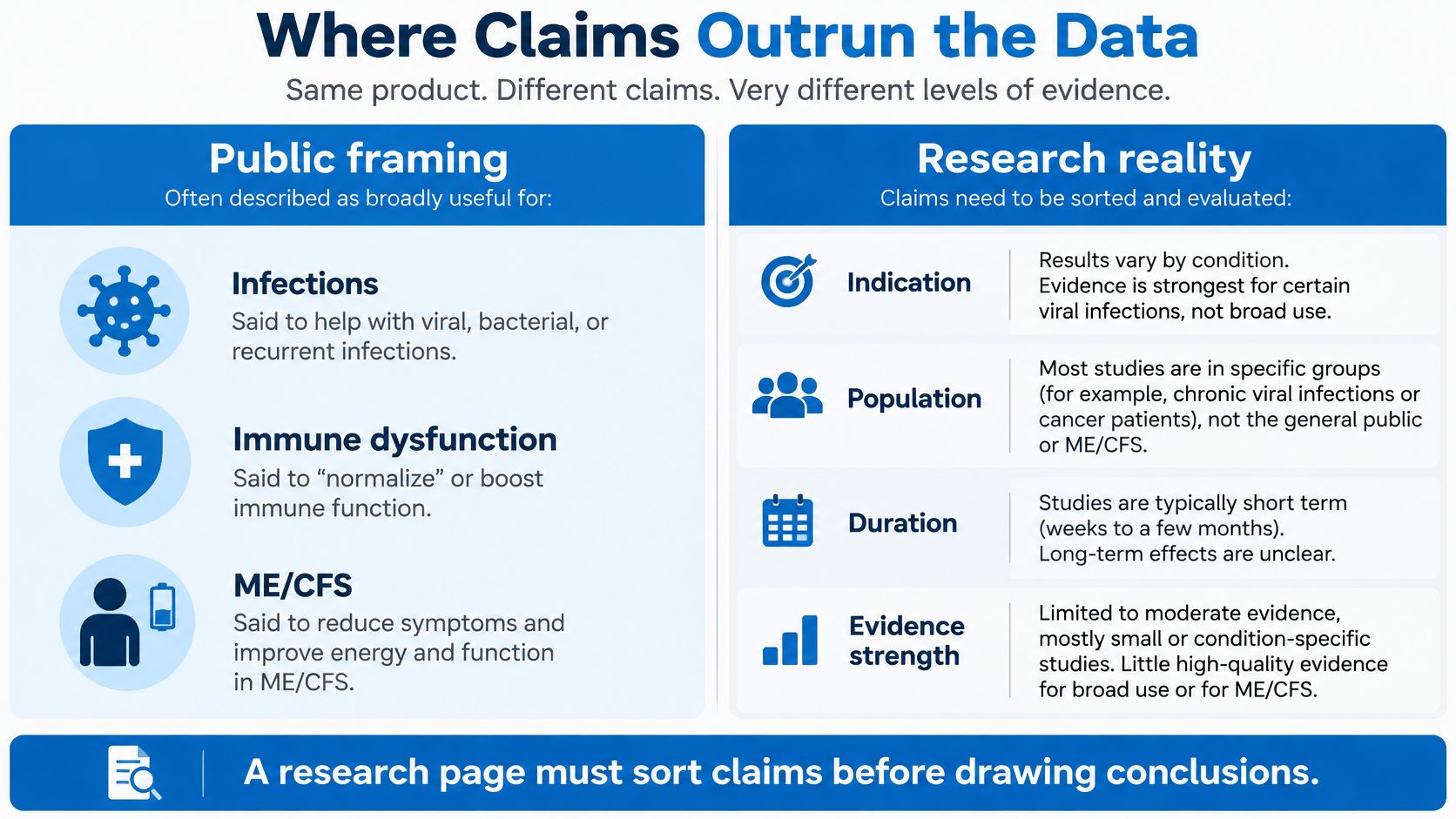
Public framing often presents Tα1 as broadly useful for infections, immune dysfunction, or ME/CFS. That framing collapses indication, population, duration, and endpoint into one promise. The comparison image below shows the sorting step a research page needs before any claim is repeated.
Claims Versus the Evidence Boundary
| Claim | Evidence type | What holds up | Boundary |
|---|---|---|---|
| Coordinates innate and adaptive immunity | Reviews plus dendritic-cell experiments | TLR2/TLR9, maturation, cytokines, IDO and regulatory T cells | Context-dependent; not a clinical outcome |
| Improves sepsis survival | RCTs, one meta-analysis, one phase 3 trial | Pooled OR favored Tα1; phase 3 did not | Investigational, not an established sepsis therapy |
| Changes pancreatitis immune markers | Five-trial meta-analysis | CD4 percentage and CD4/CD8 ratio increased | Biomarker result; adverse-event data were thin |
| Helps ME/CFS | FDA-reviewed effectiveness check | No supporting effectiveness data found | Don't infer benefit from tolerability talk |
| Is broadly safe for any population | Adult study safety summaries | No major attributable signal reported in reviewed adult studies | Not proof for pediatric, pregnancy, long-term, or combination settings |
The table separates three questions that promotional writing often merges. Can the compound affect immune cells under defined conditions? The mechanistic literature says yes, within context. Did disease-specific trials show biomarker movement? In severe acute pancreatitis, pooled trials did. Did those findings prove better clinical outcomes across conditions? No, and sepsis shows why that leap is unsafe editorially and scientifically.
ME/CFS is the clearest example. The FDA-reviewed material found no supporting effectiveness data for ME/CFS. That statement should end any implication of benefit for that indication. A compound can have an interesting immune mechanism and still have no demonstrated effectiveness for a complex condition whose endpoints weren't established in adequate trials.
Tolerability needs the same discipline. FDA-reviewed material reports that clinical studies generally found no major treatment-attributable adverse-event signal in studied adult settings, while pediatric safety has not been established. That limited statement doesn't establish safety for prolonged, repeated, or combination exposure. It also doesn't establish safety in autoimmune disease, pregnancy, immunosuppression, or childhood. Absence of a major signal in selected adult studies isn't a universal safety finding.
Pancreatitis illustrates a related gap. Immune-marker and CRP changes were reported in pooled trials, but adverse-event data in the included trials were insufficient. A favorable biomarker without adequate harm reporting gives an incomplete benefit-risk picture. Research writers should say both parts in the same paragraph, not publish the marker change and omit the missing safety record.
Several evidence gaps still need direct answers before stronger language is justified.
- Interactions: Combination settings may alter immune signaling, yet interaction data remain incomplete.
- Overstimulation: A modulator can theoretically produce unwanted immune activation in susceptible contexts, and that risk needs condition-specific testing.
- Long-term outcomes: Short trial follow-up can't establish durability or delayed harm.
- Product comparability: One batch can't be assumed equivalent to another without identity and purity documentation.
For laboratories, that last point is immediate. Identity, purity, sterility, endotoxin testing, storage conditions, and complete documentation decide whether a result can be reproduced. Claims about the peptide mean little if the material in the vial hasn't been qualified.
Laboratory Checks Before an Experiment
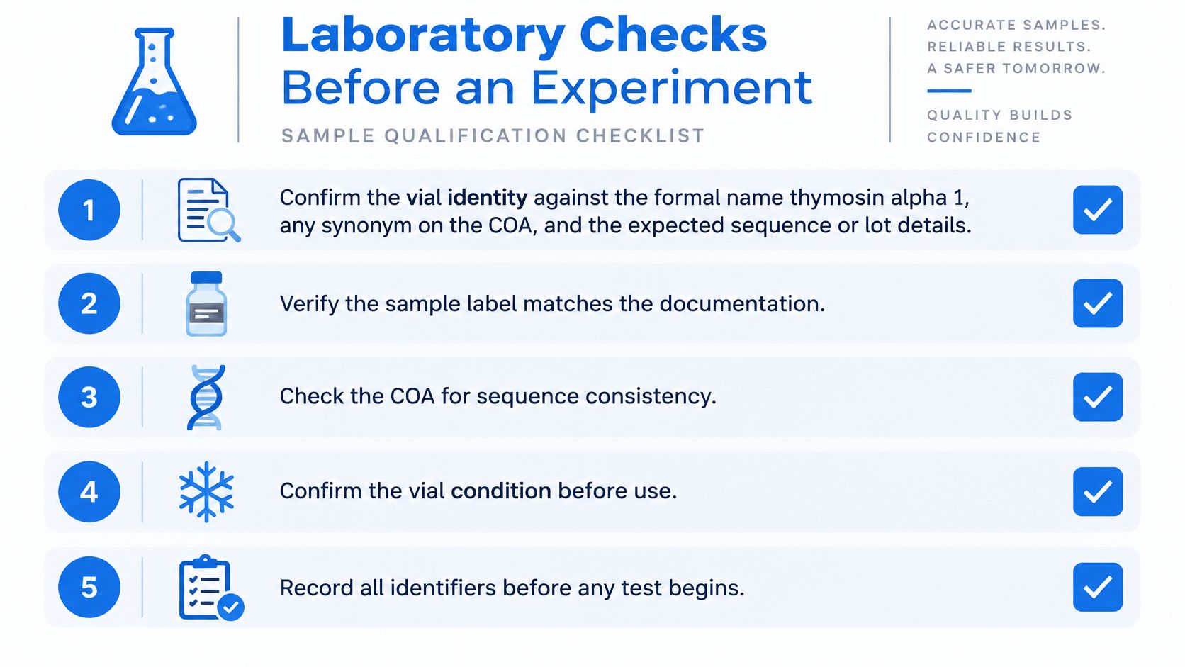
Sample qualification comes before biology. An immune assay is especially vulnerable to mistaken identity and contamination, because endotoxin and other impurities can create signals that resemble immune activity. The checklist graphic below summarizes the documentation mindset, while the numbered steps give the laboratory sequence.
A Sample-Qualification Sequence
- Confirm vial identity: Match the formal name thymosin alpha 1, any synonym on the certificate of analysis, and the expected sequence or mass for that specific lot. A similar thymic peptide name isn't close enough for a mechanistic assay.
- Read the batch COA: A generic certificate doesn't qualify the material on the bench. The batch document should state identity, purity, net content, and the analytical method used for each result.
- Check contaminant results for cell work: Sterility, endotoxin, and heavy-metal findings matter because contaminants can mimic, amplify, or mask an immune signal. A dendritic-cell assay without endotoxin information carries an avoidable interpretation risk.
- Record formulation and solubility as chemistry: Research material is commonly handled as a lyophilized solid, and supplier solubility notes guide buffer choice for sample preparation. That information belongs in the analytical or in-vitro protocol, not in any administration narrative.
- Follow specified storage conditions: Unopened material should be stored as the COA or product specification directs, typically frozen for research peptides. Freeze-thaw limits and any excursion should be entered in the laboratory notebook.
- Aliquot and label for traceability: Each aliquot needs the compound name, lot number, open date, concentration or amount prepared for the assay, and storage location. The COA should stay linked to the protocol record.
- Investigate unusual stimulation before attribution: An unexpectedly strong cytokine or maturation signal should trigger repeat contaminant checks before the result is attributed to Tα1. Endotoxin is a classic confounder in immune-cell systems.
A purity specification also needs precise language. A minimum near 98 percent, when stated on a batch document, describes the proportion assigned to the intended compound under the reported method. It doesn't promise a biological effect, and it doesn't remove the need for identity confirmation. Purity, identity, and activity are separate questions.
Laboratory rule: If another researcher can't identify the exact lot, method, storage history, and contaminant status from the record, the experiment isn't yet reproducible.
Documentation should travel with the data. Figures for dendritic-cell maturation, T-cell subsets, or cytokine panels should be traceable to the lot and preparation record used on that day. When a repeat run uses a new lot, comparability should be checked rather than assumed.
The permitted frame is in-vitro investigation, analytical comparison, stability assessment, and method development. Those activities can test mechanism and material quality without converting literature findings into human or veterinary claims.
Research Supply Limits and the Required Disclaimer
Procurement has a boundary as clear as the evidence boundary. Pro Tec Pep supplies research compounds for analytical testing and lawful nonclinical work. Products are sold strictly for research use and aren't for human or veterinary use, diagnosis, treatment, or prevention. Pro Tec Pep products aren't intended for consumption or administration, and the supplier doesn't recommend products for personal, therapeutic, diagnostic, or veterinary purposes.
None of Pro Tec Pep's products or statements have been evaluated by the FDA. A catalog listing therefore doesn't make a compound a medicine, and a research peptide isn't interchangeable with a prescription product or supplement. Literature on Tα1 may inform assay design, but it doesn't change the research-only status of supplied material.
Permitted research includes analytical testing, chemical characterization, method development, reference-standard comparison, stability testing, instrument calibration, and in-vitro laboratory investigation, along with other lawful nonclinical activities. Pro Tec Pep doesn't provide administration, treatment, or medical-use guidance. Nothing on the Pro Tec Pep website is medical advice.
Documentation at Procurement
Each listed research-peptide page links a certificate of analysis, and that certificate is the document researchers should match to the purchased lot before an assay. The Research Peptide category has 22 results, with 12 shown per page, so product-level documentation matters more than a category-level assumption. A COA for one lot shouldn't be used to qualify another.
Identity, purity, sterility, endotoxin status, storage history, and the COA still decide whether a run can be reproduced. Those checks are not administrative decoration. In immune-cell research, they're part of the scientific result because poor material control can manufacture a false pathway signal or erase a real one.
For laboratory research use only. Not for human or veterinary use. Not a drug, food, or cosmetic. Not evaluated by the FDA. Not intended to diagnose, treat, cure, or prevent any disease. By purchasing, you certify you are a qualified researcher and agree to use this product solely for in-vitro laboratory research.
Researchers planning in-vitro work on thymosin alpha 1 signaling can review batch documentation and research-only compound listings through Pro Tec Pep. The catalog is built for analytical testing, chemical characterization, and lawful nonclinical investigation, with product-level COA access that supports lot-matched experimental records.